
Figure 2.
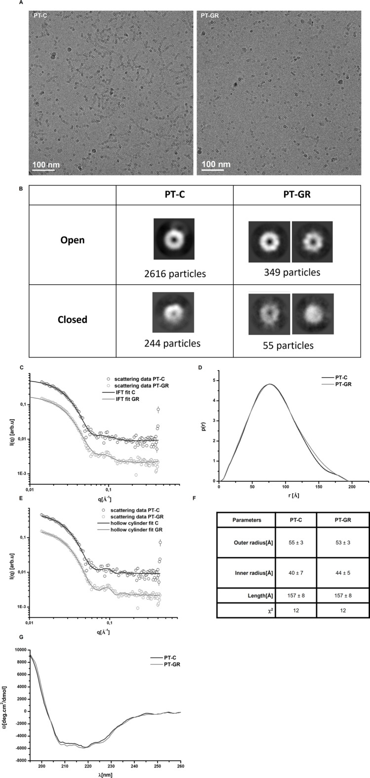
Glucose restriction does not change the conformation of the 20S proteasome. (A) Representative cryo-transmission electron microcopy (cryo-TEM) micrographs of 20S proteasome particles isolated from cells cultured under control (PT-C) or glucose restriction (PT-GR) conditions. (B) 2D classification of pore opening and frequency distribution in experimental samples performed with cryoSPARC software. (C–F) PT-C or PT-GR samples were analyzed by SAXS as described in experimental procedures. (C) Scattering curves, (D) distance pair distribution function, p(r), (E) fitting of scattering curves using a form factor of hollow cylinder, and (F) hollow cylinder modelling results. (G) Circular dichroism curves obtained for PT-C or PT-GR samples as described in experimental procedures. The data is representative of 2 independent experiments.