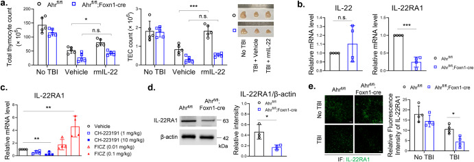
Fig. 2. Knockout of AHR in TECs decreased IL-22RA1 expression.
a B6-Ahrfl/fl mice and B6-Ahrfl/fl;Foxn1-cre mice, pretreated by 5.5 Gy TBI, were intraperitoneally injected with vehicle or rmIL-22 (200 ng) thrice weekly for 2 weeks. At day 14, thymus cells were counted as described (n = 5). b, c Thymii were collected from irradiated mice. qPCR was performed to detect mRNA levels of IL-22 and IL-22RA1 (n = 4). The values of control groups were set as 1, and the values of other groups were 2–ΔΔCT relative to controls. d Thymic stromal cells were isolated from irradiated mice (n = 3). Western blot was performed with indicated antibodies. Band intensity was analyzed using ImageJ. e Cryosections of thymii were stained by immunofluorescence with anti-IL-22RA1 (n = 4). Fluorescence intensity was analyzed using ImageJ. Scale bar: 50 μm. Data are mean ± SD, compared using one-way ANOVA test or Student’s t test. *, p < 0.05; **, p < 0.01; ***, p < 0.001; n.s., not significant.

