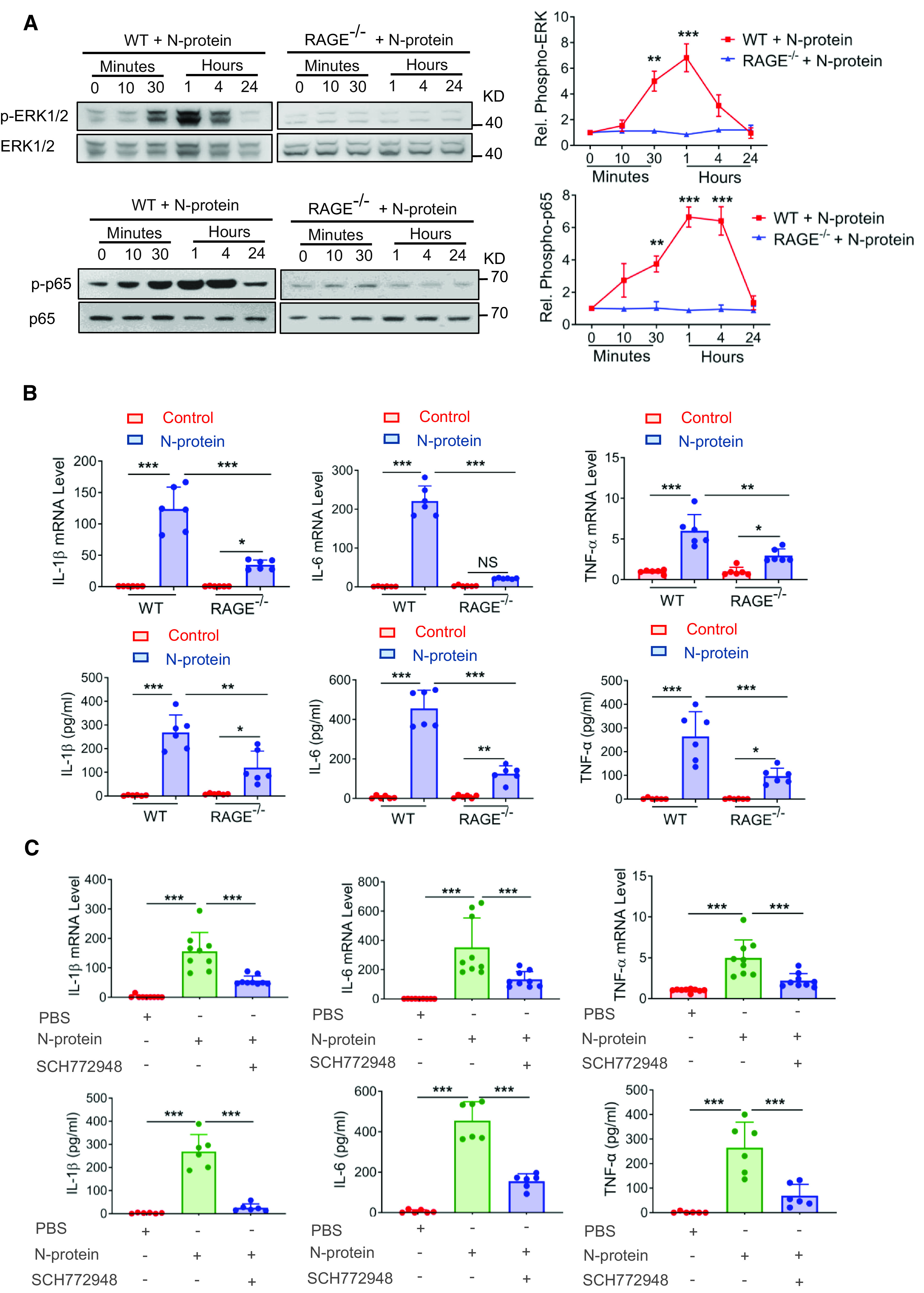
Figure 4.

RAGE knockout reduced inflammatory response induced by N-protein in BMDMs. (A) BMDMs from WT and RAGE knockout mice were treated with recombinant full-length N-protein (5 μg/ml) for the indicated times. Cell lysate was analyzed via Western blot using antibodies specific to phosphorylated (upper panels) or total (lower panels) of ERK1/2 or NF-ĸB p65. The levels of phosphorylation were quantified with ImageJ software (n = 3). (B) BMDMs from WT and RAGE knockout mice were treated with N-protein (5 μg/ml) for 24 hours. The mRNA expression of proinflammatory cytokines IL-1β, IL-6, and TNF-α in BMDMs was quantitated by qRT-PCR (upper panel, n = 6). Culture supernatants were analyzed by ELISA for IL-1β, IL-6, and TNF-α (lower panel, n = 6). (C) BMDMs from WT mice were pretreated with ERK1/2 inhibitor (SCH772948, 500 nM) for 1 hour and then stimulated with recombinant N-protein (5 μg/ml) for 24 hours. The mRNA expression of proinflammatory cytokines IL-1β, IL-6, and TNF-α was quantitated by qRT-PCR (upper panel, n = 9). Culture supernatants were analyzed by ELISA for IL-1β, IL-6, and TNF-α (lower panel, n = 6). Data are presented as mean ± SD. *P < 0.05, **P < 0.01, and ***P < 0.001. One-way ANOVA with Tukey’s post hoc test versus time 0 (A). Two-way ANOVA with Tukey’s post hoc test (B). One-way ANOVA with Tukey’s post hoc test (C).
