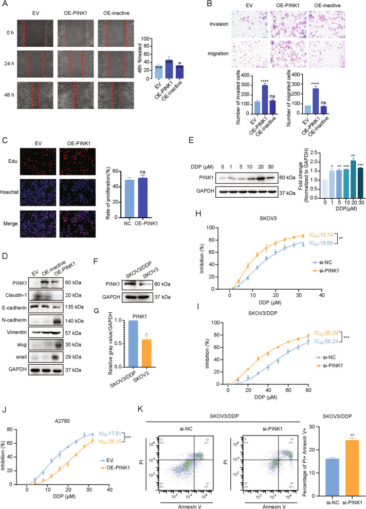
Fig. 2.
PINK1 promotes metastasis and chemoresistance of ovarian cancer in vitro. A Cells were transfected with over-expression plasmid for 24 h. Representative images of the wound scratch assay utilizing the A2780 cell lines after scratching 24 and 48 h. The histograms on the right show the quantitative results of the healing percentage after 48 h of three independent replicates. B Effects of PINK1 on A2780 cell invasion (upper) and migration (bottom). Cells were transfected by PINK1 over-expression plasmid for 24 h and then measured using transwell assay with (invasion) or without (migration) Matrigel after incubation for 24 h. Invasion or migration cells were fixed, stained, photographed, and counted in 6 random views. The quantitative analysis of invade or migrated cells were shown on the bottom. C Representative images (left) and quantitative analysis (right) of proliferating A2780, which were transfected by Flag-PINK1 over-expression vector for 48 h and then assessed by EdU kit assay. D Western blot analysis of EMT markers. A2780 cells were transfected by PINK1 over-expression plasmid. E The protein (left) and mRNA (right) levels of PINK1 in ovarian cancer cells treated with DDP as indicated concentrations for 24 h. F and G The protein levels of PINK1 in SKOV3 cell and SKOV3/DDP cell by western blot analysis (F) and the quantification analysis (G). H and I IC50 values of cisplatin are tested in PINK1 targeting siRNA-transfected SKOV3 cells (H) and SKOV3/DDP cells (I) by CCK-8 assay. J IC50 values of cisplatin are tested in PINK1 over-expressed A2780 cells. Results were shown as mean ± standard deviation (SD) in H-J. K FCM analysis on SKOV3/DDP cells treated with PINK1 targeting siRNA and cisplatin (20μM) for 48 h. ns, no statistical significance. * p < 0.05, ** p < 0.01, *** p < 0.001, **** p < 0.0001. Values are mean ± SEM. Data are representative of 3 independent experiments

