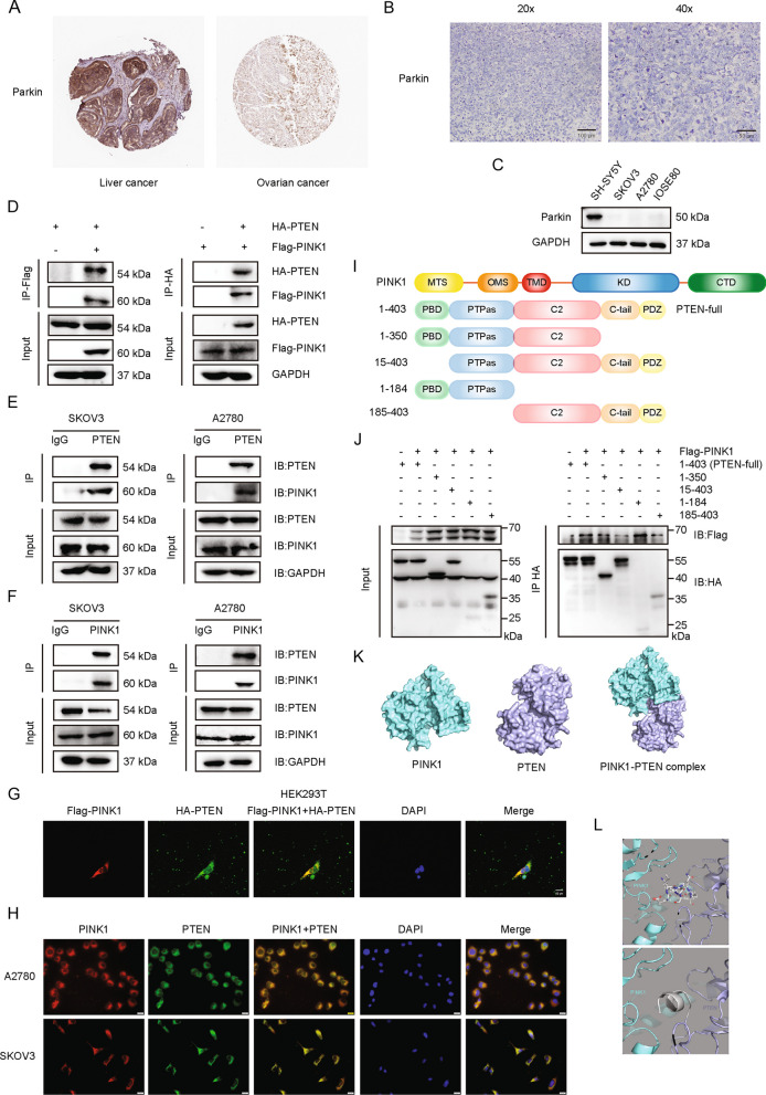
Fig. 3.
PINK1 interacts with PTEN. A Representative IHC image of Parkin from ovarian cancer patient and liver cancer patient downloaded from The Human Protein Atlas. B Representative IHC image of Parkin from local ovarian cancer patient. C Western blot for Parkin expression in four cell lines. D Immunoblotting analysis of Flag-PINK1 and HA-PTEN expression in a co-IP assay performed using Protein A/G PLUS-Agarose and anti-Flag (left) or anti-HA (right) primary antibody in HEK293T cells. E and F Immunoblotting analysis of endogenous PINK1 and PTEN expression in a co-IP assay performed in A2780 and SKOV3 cells with Protein A/G PLUS-Agarose and anti-PTEN (E) or anti-PINK1 (F) primary antibody. G Confocal microscopy detection of the co-localization of Flag-PINK1 (red) and HA-PTEN (green) in HEK293T cells. Nuclei were stained using DAPI (blue). DAPI, 4’,6-diamidino-2-phenylindole. H Immunofluorescence assay of the co-localization of endogenous PINK1 (red) and PTEN (green) in A2780 and SKOV3 cells. I Schematic diagram of PINK1 and truncated structure of PTEN. PBD, phosphatidylinositol (PtdIns) (4,5)P2-binding domain; PTPas, phosphatase domain; C2, C2 lipid/membrane-binding domain; C-tail, containing Pro, Glu, Ser and Thr (PEST) sequences; PDZ, PDZ-binding motif. MTS, mitochondrial targeting sequence; OMS, outer membrane localization signal; TMD, transmembrane domain; KD, kinase domain; CTD, C-terminal domain. J Flag-PINK1 was expressed with HA-PTEN (full length) or PTEN deletion mutants in HEK293T cells. Total cell lysates were subjected to HA affinity purification and analyzed by immunoblotting with anit-Flag and anti-HA antibodies. K Molecular docking model of PINK1/PTEN interaction by Z-DOCK. L Diagram of binding mode of PINK1 (green) and PTEN (purple) predicted by Z-docking server. The predicted binding sites are shown in stick mode in the upper panel and in cartoon mode in the bottom panel (grey). PTEN residues 1–14 are located at the binding interface of two proteins and may form contact with PINK1. Data are representative of 3 independent experiments

