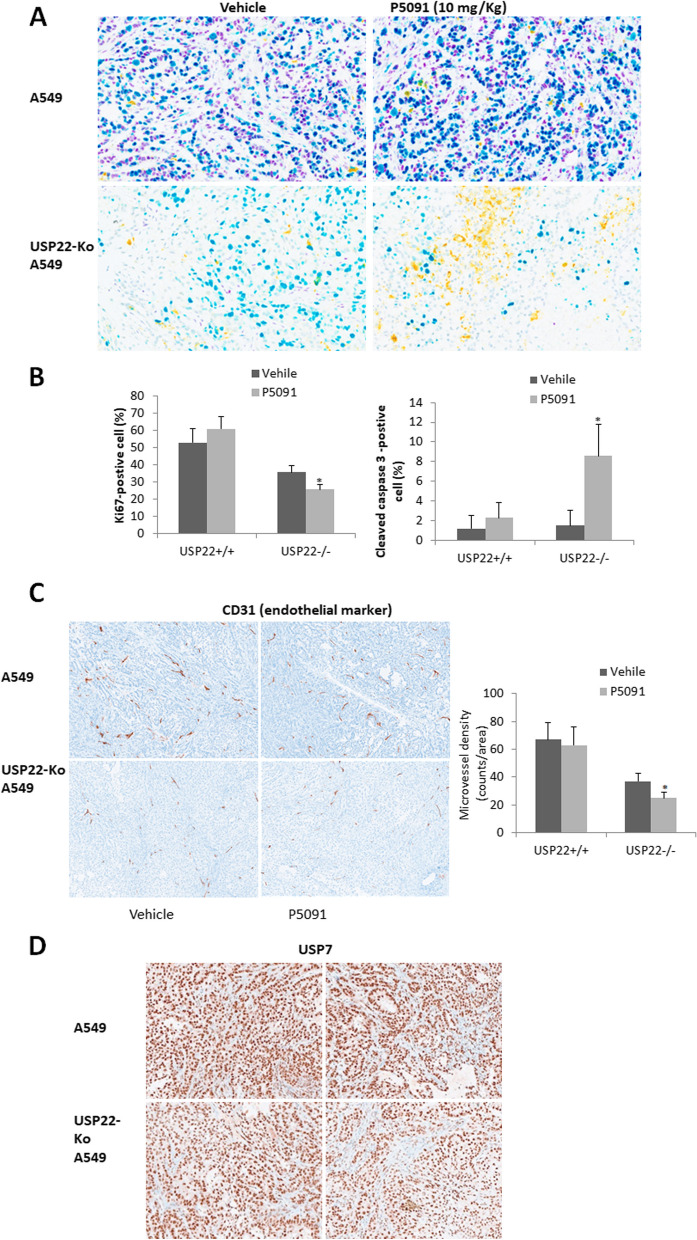
Fig. 7.
PD5091 induced more apoptosis and suppressed angiogenesis in USP22-Ko cancer cells. A. Multiplex IHC analysis of USP22 (Purple), Ki67 (Teal), and cleaved caspase-3 (Yellow) in the parental and USP22-Ko A549 xenograft tissues treated with P5091. B. Semi-quantitative analysis of Ki67-positive and cleaved caspase 3- positive cell populations in the parental and USP22-Ko A549 treated with P5091. C. IHC analysis of CD31 (Brown) in the parental and USP22-Ko A549 xenograft tissues treated with P5091 (Left panel), and semi-quantitative analysis of MVD in these xenografts (Right panel), *p<0.05, compared to Vehicle. D. IHC analysis of USP7 (Brown) in the parental and USP22-Ko A549 xenografts treated with P5091

