Abstract
OBJECTIVE--To study the role of magnetic resonance imaging (MRI) in evaluating children with shunted hydrocephalus. METHODS--Sixty one asymptomatic children with shunted hydrocephalus or cystic cerebrospinal fluid collections were studied by cranial MRI. The information obtained from the images was classified into three categories: provided (1) a new diagnosis, (2) additional information, or (3) no essential new information. The findings were compared with those of the last follow up computed tomograms. RESULTS--MRI provided a new diagnosis in seven cases (11.5%), and additional information was obtained in 34 (55.7%) cases. In 20 cases (32.8%) no essential new information was obtained. MRI visualised white matter lesions and corpus callosum pathology more often than computed tomograms. CONCLUSIONS--MRI provided new important information in cases of children with shunted hydrocephalus to such an extent that it can be recommended as the primary imaging method for every child with this disorder.
Full text
PDF
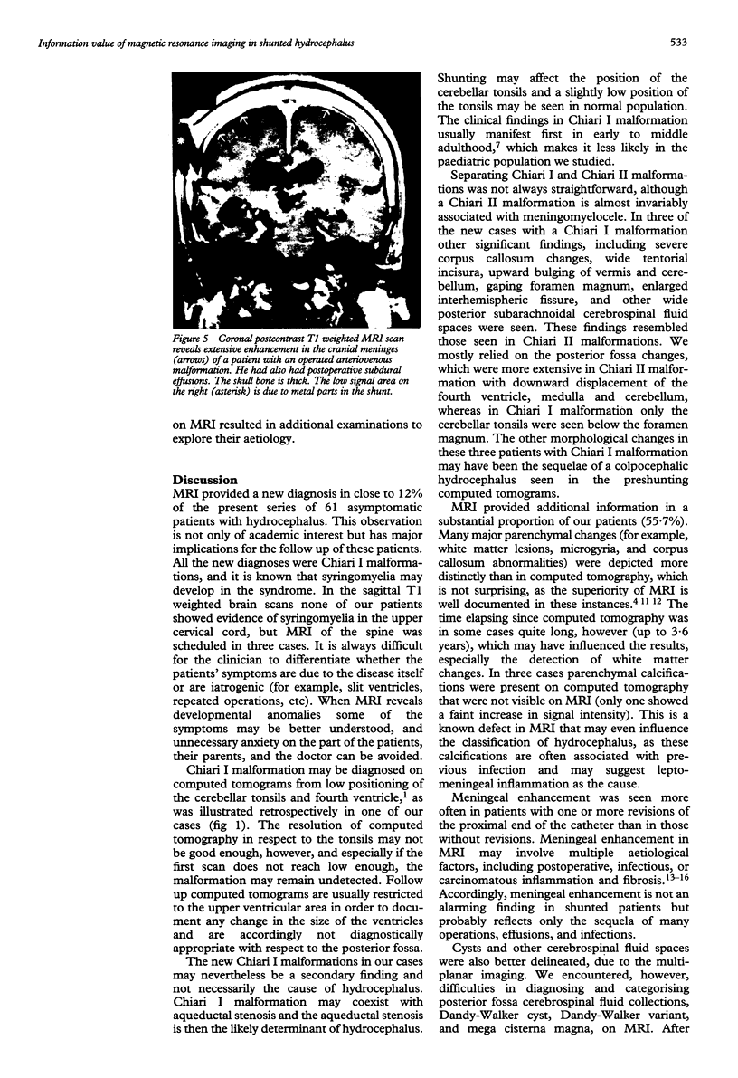
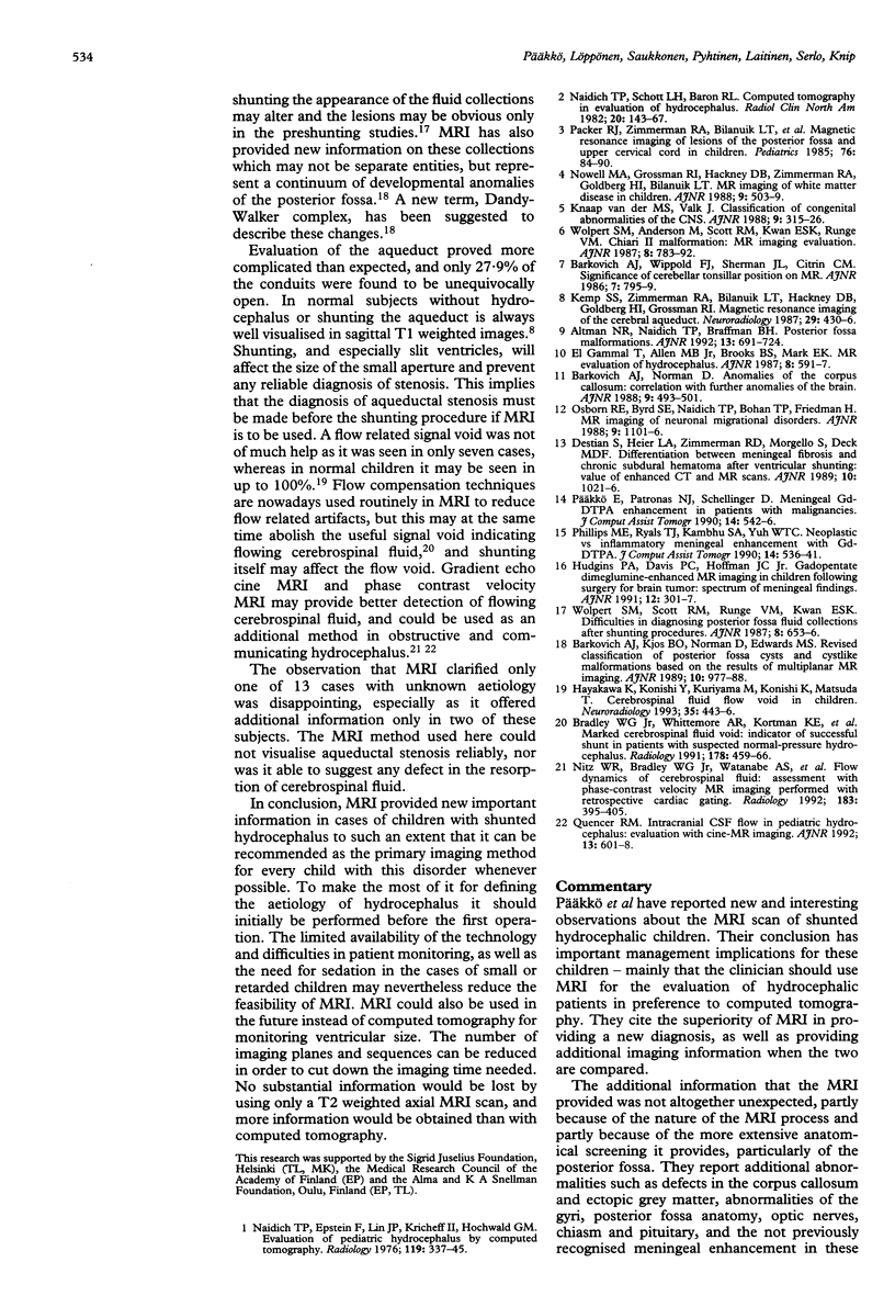
Images in this article
Selected References
These references are in PubMed. This may not be the complete list of references from this article.
- Altman N. R., Naidich T. P., Braffman B. H. Posterior fossa malformations. AJNR Am J Neuroradiol. 1992 Mar-Apr;13(2):691–724. [PMC free article] [PubMed] [Google Scholar]
- Barkovich A. J., Wippold F. J., Sherman J. L., Citrin C. M. Significance of cerebellar tonsillar position on MR. AJNR Am J Neuroradiol. 1986 Sep-Oct;7(5):795–799. [PMC free article] [PubMed] [Google Scholar]
- Bradley W. G., Jr, Whittemore A. R., Kortman K. E., Watanabe A. S., Homyak M., Teresi L. M., Davis S. J. Marked cerebrospinal fluid void: indicator of successful shunt in patients with suspected normal-pressure hydrocephalus. Radiology. 1991 Feb;178(2):459–466. doi: 10.1148/radiology.178.2.1987609. [DOI] [PubMed] [Google Scholar]
- Destian S., Heier L. A., Zimmerman R. D., Morgello S., Deck M. D. Differentiation between meningeal fibrosis and chronic subdural hematoma after ventricular shunting: value of enhanced CT and MR scans. AJNR Am J Neuroradiol. 1989 Sep-Oct;10(5):1021–1026. [PMC free article] [PubMed] [Google Scholar]
- Hayakawa K., Konishi Y., Kuriyama M., Konishi K., Matsuda T. Cerebrospinal fluid flow void in children. Neuroradiology. 1993;35(6):443–446. doi: 10.1007/BF00602825. [DOI] [PubMed] [Google Scholar]
- Hudgins P. A., Davis P. C., Hoffman J. C., Jr Gadopentetate dimeglumine-enhanced MR imaging in children following surgery for brain tumor: spectrum of meningeal findings. AJNR Am J Neuroradiol. 1991 Mar-Apr;12(2):301–307. [PMC free article] [PubMed] [Google Scholar]
- Kemp S. S., Zimmerman R. A., Bilaniuk L. T., Hackney D. B., Goldberg H. I., Grossman R. I. Magnetic resonance imaging of the cerebral aqueduct. Neuroradiology. 1987;29(5):430–436. doi: 10.1007/BF00341738. [DOI] [PubMed] [Google Scholar]
- Naidich T. P., Epstein F., Lin J. P., Kricheff I. I., Hochwald G. M. Evaluation of pediatric hydrocephalus by computed tomography. Radiology. 1976 May;119(2):337–345. doi: 10.1148/119.2.337. [DOI] [PubMed] [Google Scholar]
- Naidich T. P., Schott L. H., Baron R. L. Computed tomography in evaluation of hydrocephalus. Radiol Clin North Am. 1982 Mar;20(1):143–167. [PubMed] [Google Scholar]
- Nitz W. R., Bradley W. G., Jr, Watanabe A. S., Lee R. R., Burgoyne B., O'Sullivan R. M., Herbst M. D. Flow dynamics of cerebrospinal fluid: assessment with phase-contrast velocity MR imaging performed with retrospective cardiac gating. Radiology. 1992 May;183(2):395–405. doi: 10.1148/radiology.183.2.1561340. [DOI] [PubMed] [Google Scholar]
- Osborn R. E., Byrd S. E., Naidich T. P., Bohan T. P., Friedman H. MR imaging of neuronal migrational disorders. AJNR Am J Neuroradiol. 1988 Nov-Dec;9(6):1101–1106. [PMC free article] [PubMed] [Google Scholar]
- Paakko E., Patronas N. J., Schellinger D. Meningeal Gd-DTPA enhancement in patients with malignancies. J Comput Assist Tomogr. 1990 Jul-Aug;14(4):542–546. doi: 10.1097/00004728-199007000-00008. [DOI] [PubMed] [Google Scholar]
- Packer R. J., Zimmerman R. A., Bilanuik L. T., Leurssen T. G., Sutton L. N., Bruce D. A., Schut L. Magnetic resonance imaging of lesions of the posterior fossa and upper cervical cord in childhood. Pediatrics. 1985 Jul;76(1):84–90. [PubMed] [Google Scholar]
- Phillips M. E., Ryals T. J., Kambhu S. A., Yuh W. T. Neoplastic vs inflammatory meningeal enhancement with Gd-DTPA. J Comput Assist Tomogr. 1990 Jul-Aug;14(4):536–541. doi: 10.1097/00004728-199007000-00007. [DOI] [PubMed] [Google Scholar]
- Quencer R. M. Intracranial CSF flow in pediatric hydrocephalus: evaluation with cine-MR imaging. AJNR Am J Neuroradiol. 1992 Mar-Apr;13(2):601–608. [PMC free article] [PubMed] [Google Scholar]
- Wolpert S. M., Scott R. M., Runge V. M., Kwan E. S. Difficulties in diagnosing congenital posterior fossa fluid collections after shunting procedures. AJNR Am J Neuroradiol. 1987 Jul-Aug;8(4):653–656. [PMC free article] [PubMed] [Google Scholar]
- van der Knaap M. S., Valk J. Classification of congenital abnormalities of the CNS. AJNR Am J Neuroradiol. 1988 Mar-Apr;9(2):315–326. [PMC free article] [PubMed] [Google Scholar]


