Fig. 1.
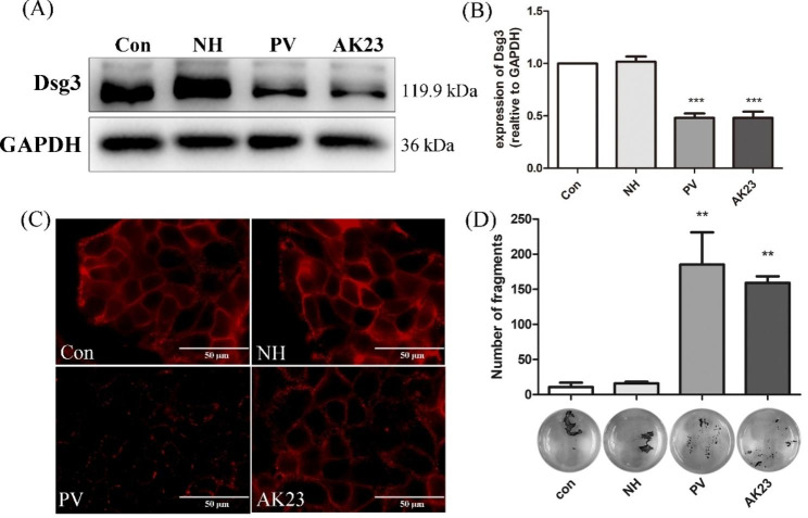
Both pemphigus sera and AK23 caused a decrease in keratinocyte adhesion and Desmoglein (Dsg) 3 internalization in HaCaT cells. (A) Western blot analysis of Dsg3 in total cell lysates of HaCaT cells incubated with 5% PV sera or 1 µg/ml AK23 for 24 h. GAPDH was used as a loading control. Respective controls [normal control group (Con) or normal healthy sera group (NH)] were run in parallel. Both PV sera and AK23 decreased Dsg3 protein levels as compared with the control. (B) Densitometric analysis was derived from the ratio of Dsg3 to GAPDH expression in each group and then normalized to the control group. Statistically significant differences are indicated by *** p < 0.001 vs. Con. Data are represented by mean ± SE (n = 3). (C) Immunostaining revealed that a 24-h incubation with 5% PV sera or 1 µg/ml AK23 induced fragmentation of the Dsg3 staining pattern along the cell borders (scale bar = 50 μm). (D) Dissociation assays in HaCaT cells showed weaker intercellular adhesion consistence with more free cells in cells incubated with 5% PV sera or AK23. ** p < 0.01 vs. Con. Data are shown as mean ± SE (n = 3)

