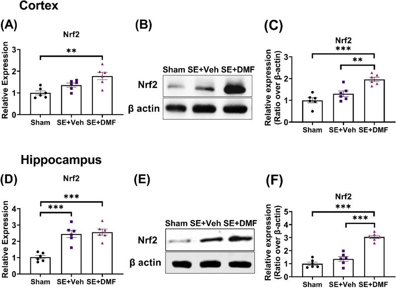
Fig. 1.
DMF increased Nrf2 expression in the cortex and hippocampus after SE. A, D Relative gene expression of Nrf2 measured by quantitative Reverse transcription-polymerase chain reaction (RT-PCR) in the cortex (A) and the hippocampus (D) of control rats (n = 6), and rats after kainic acid-induced SE (KA-SE) treated with either vehicle (n = 6) or DMF (ip, 200 mg/kg/day, 7 days, n = 6). B, E Representative western blot images of Nrf2 in the cortex (B) and the hippocampus (E). C, F Quantification of western blotting results for Nrf2 in the cortex (C; n = 6) and hippocampus (F; n = 6). Gene expression was normalized to GAPDH expression and is presented as fold-change compared to the level in sham rats. Protein levels were expressed as relative protein expression normalized to β-actin. Data are reported as the mean ± SEM. **p < 0.01 and ***p < 0.001 after one-way ANOVA followed by Tukey’s posthoc test

