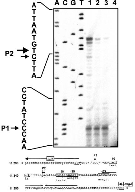
FIG. 5.
Mapping of the 5′ start site of ppk mRNA by primer extension. (Upper panel) Autoradiograph. RNA was prepared from ADP1 cells growing in minimal medium with 100 μM (lane 1) or 1 mM (lane 2) phosphate or growing exponentially in LB medium (lane 3). The primer extension control without RNA is also shown (lane 4). Lanes A, C, G, and T show the sequencing products obtained with the same primer. The sequence is shown on the left. Arrows indicate the start sites of transcription (P1 and P2). (Lower panel) Sequence interpretation. The sequence of the coding strand upstream of ppk is shown. The nucleotide positions are given on the left in accordance with the diagram shown in Fig. 1. The start codon of ppk and stop codon of mtgA are underlined, and the direction of translation is indicated by arrows. The putative transcriptional terminator sequence of mtgA is marked by inverted arrows, and the bases matching the inverted repeat are indicated by uppercase letters. The start sites of transcription (P1 and P2) are indicated by downward arrows, and the sequences with the highest similarity to E. coli ς70 promoter sequences (−10, −35) are boxed.

