
Figure 4. Mast cell deficiency reduces GFAP expression and alters astrocyte morphology in 5XFAD mice.
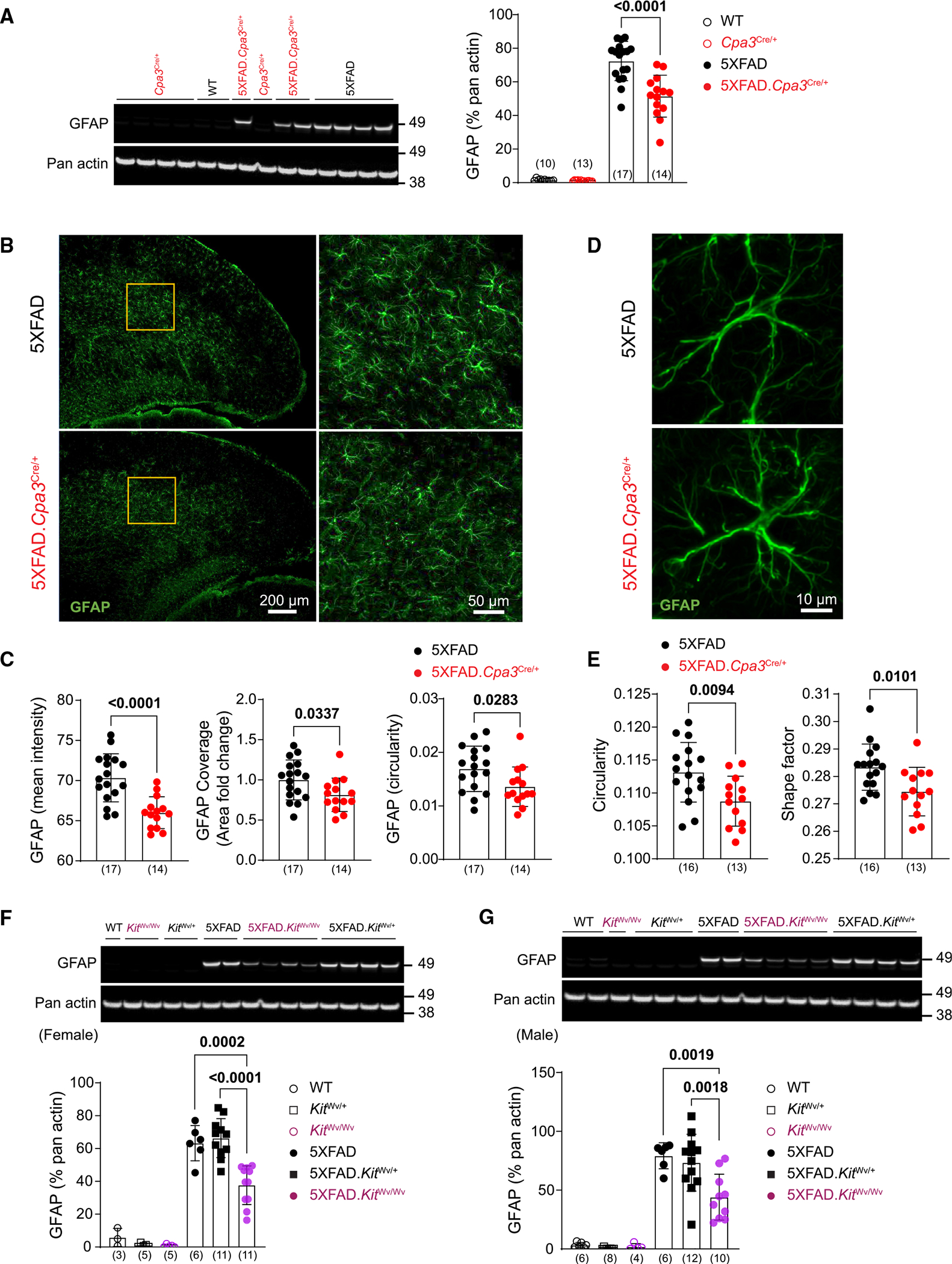
(A) Representative western blots and protein levels for reactive astrocyte marker GFAP measured from cortical homogenates of 8- to 9-month-old female Cpa3Cre cohort mice.
(B) Brain cortical sections were labeled with GFAP (green) antibody. Representative images of cortex from 8- to 9-month-old female 5XFAD and 5XFAD.Cpa3Cre/+ mice. The details of GFAP-positive astrocytes in the inserts are shown.
(C) Quantification of mean fluorescence intensity, area fold change, and circularity of GFAP-positive cells in (B).
(D) Representative images of a single astrocyte in the visual cortex of 8- to 9-month-old female 5XFAD and 5XFAD.Cpa3Cre/+ mice.
(E) Quantification of GFAP-positive astrocyte circularity and shape factor in (D).
(F and G) Representative western blots and protein levels for astrogliosis marker GFAP measured from cortical homogenates of 7- to 9-month-old female (F) or 8- to 10-month-old male (G) KitWv cohort mice. GFAP, glial fibrillary acidic protein. Bar graphs depict mean ± SD with individual mice as points. The number of mice is specified in the panels. Two-way ANOVA with Tukey’s multiple comparison tests (A, F, and G) and unpaired t test (C and E) were performed to determine significance. See also Figure S4.