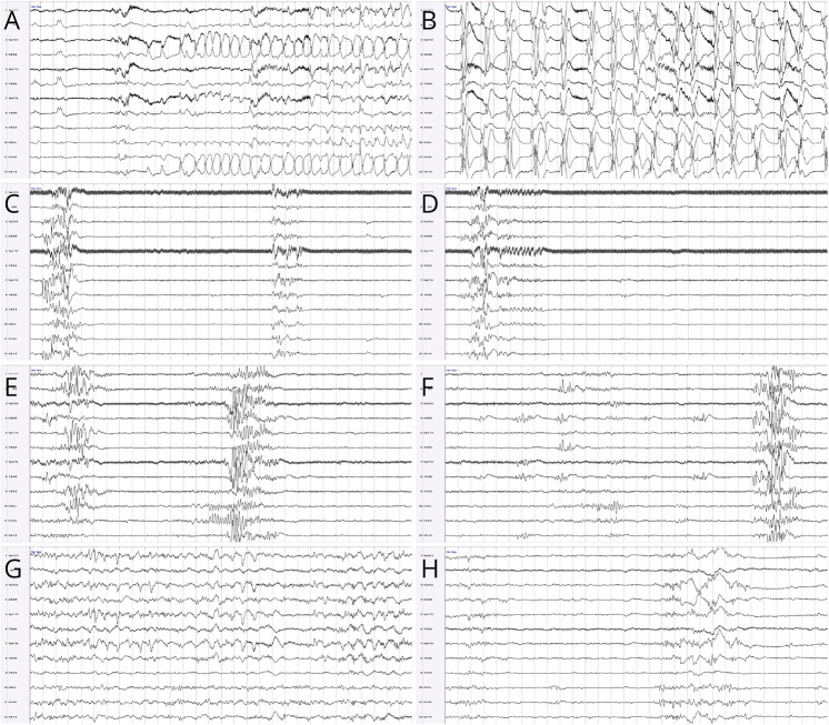
Figure 1. Evolution of EEG Findings.
Modified neonatal double distance montage, read at 15 μV for panels A and B and at 7 μV for panels D–H, with 30 seconds per page in all panels. Electrographic seizures with right centrotemporal onset (A) and evolution to rhythmic bilateral large amplitude spike-and-wave discharges (B). Background activity before the pyridoxine load, showing a severely abnormal burst-suppression pattern for 60 consecutive seconds (C, D). Background activity 48 hours after the pyridoxine load, showing an ongoing abnormal burst-suppression pattern (E), although some activity is rarely seen within the interburst interval (F). Background activity 5 days after the pyridoxine load, showing normal activity with typical neonatal electrographic elements during wakefulness (G), and mildly excessive discontinuity during quiet sleep (H).

