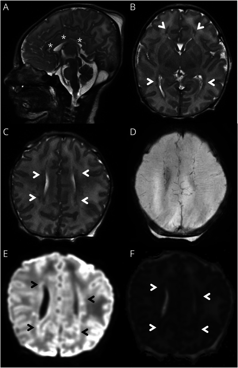
Figure 2. Brain MRI Findings.

Sagittal T2 sequence showing diffusely thin corpus callosum (*, A). Axial T2 sequence showing areas of abnormal signal in the periventricular white matter anterior to the frontal horns and posterolateral to the occipital horns (arrowheads, B). Axial T2 sequence (rostral to B) showing small multifocal areas of abnormal signal in the periventricular white matter (arrowheads, C). Corresponding SWI sequence showing absence of hemorrhage (D). Corresponding DWI (E) and ADC (F) sequencing showing areas of restricted diffusion in the periventricular white matter, consistent with acute injury (arrowheads). ADC = apparent diffusion coefficient; DWI = diffusion-weighted imaging.
