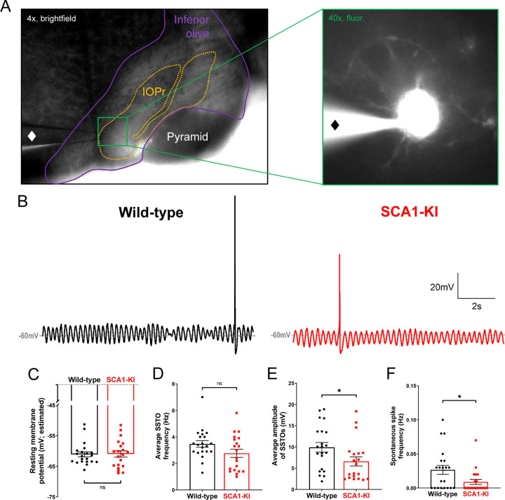
Figure 5. Spontaneous activity in SCA1-KI IOPr neurons is largely unchanged.
(A) Images demonstrating whole-cell patch-clamp electrophysiology on IO neurons in coronal brainstem slices at 4x magnification (left) and 40x magnification (right). Internal solution included a small amount of fluorescent dye that filled cells while recording, allowing for the single-cell morphological analysis shown in Fig. 1. A ♦ symbol marks the recording electrode. (B) Representative traces of spontaneous activity in IOPr neurons from wild-type (left) and SCA1-KI (right) mice at an early symptomatic timepoint of 14 weeks. The presence of spontaneous subthreshold oscillations (SSTOs) was used as confirmation of cell type when recording. (C–F) Quantification of various electrophysiological properties show few differences between SCA1-KI and wild-type IOPr neurons at rest. There was no apparent change in resting membrane potential┼ (C) or SSTO frequency (D) between genotypes, though SCA1-KI IOPr neurons did exhibit diminished SSTO amplitude (E) and spontaneous spike generation (F). Data are expressed as mean ± SEM. Statistical significance derived by unpaired t-test with Welch’s correction, * = P < 0.05, ns = not significant. ┼Due to the persistence of SSTOs, the average membrane potential across 10 s or recording was used as an estimate of resting membrane potential. Nwild-type = 21 cells from 15 mice, NSCA1-KI = 20 cells from 13 mice.

