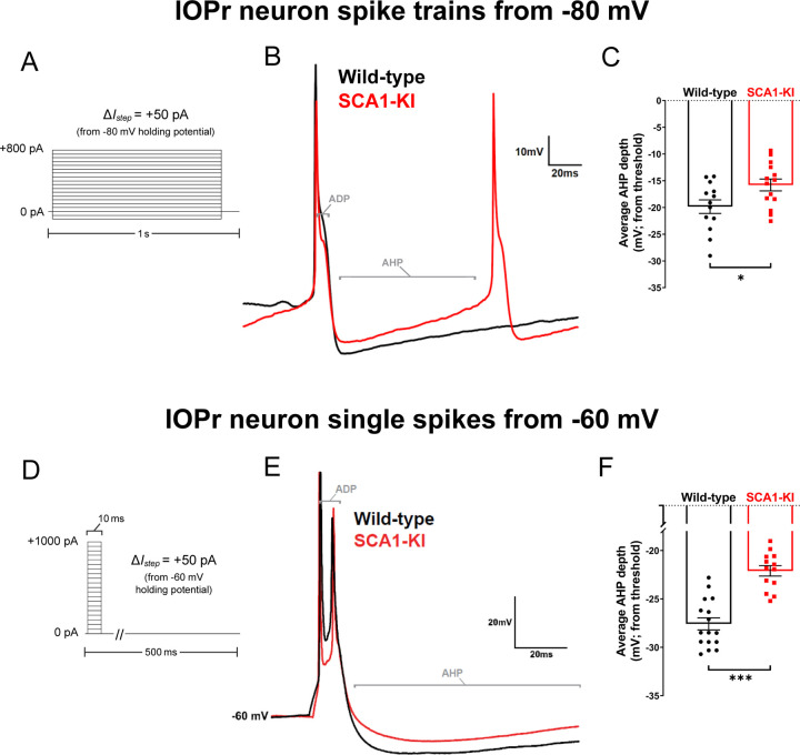
Figure 8. Spike afterhyperpolarization is diminished in SCA1-KI IOPr neurons.
(A) In order to assess the functional relevance of ion channel loss in SCA1-KI IOPr neurons, spike trains were generated by injecting increasing levels of current from −80 mV (a holding potential at which active conductances are minimal in the IO). (B) Representative traces of spikes generated by injecting +450 pA current in IOPr neurons of wild-type and SCA1-KI mice at 14 weeks. (C) Quantification of AHP depth (measured from each spike’s threshold), demonstrating that SCA1-KI IOPr spikes during spike trains exhibit a shallower AHP compared to wild-type. (D) In order to determine if this AHP loss can also occur at rest, single spikes were generated by injecting increasing levels of current from −60 mV (a holding potential close to resting Vm). Because the amount of current needed to generate a single spike varied between cells, the first spike that exhibited an ADP was used for comparisons. (E) Representative traces of evoked activity in IOPr neurons in wild-type and SCA1-KI mice at 14 weeks are overlaid. (F) Quantification of AHP depth (measured from each spike’s threshold), demonstrating that SCA1-KI IO neuron spikes from rest also exhibit a shallower AHP compared to wild-type. Data are expressed as mean ± SEM. Statistical significance derived by unpaired t-test with Welch’s correction, * = P < 0.05, *** = P < 0.001. (A–C) Nwild-type = 13 cells from 12 mice, NSCA1-KI = 14 cells from 9 mice. (D–F) Nwild-type = 16 cells from 14 mice, NSCA1-KI = 13 cells from 9 mice.

