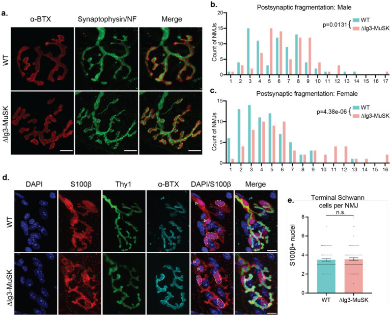
Figure 1. NMJs in young adult ΔIg3-MuSK mice are fragmented postsynaptically but maintain anatomical innervation and normal complement of terminal Schwann cells.
a. WT and ΔIg3-MuSK NMJs with postsynaptic AChRs in red and nerve terminals and axons in green, bar = 10μm. Note the increase in postsynaptic fragmentation. b. Quantification of postsynaptic fragmentation in male WT and ΔIg3-MuSK mice. c. Quantification of postsynaptic fragmentation in female WT and ΔIg3-MuSK mice. (generalized linear models, full data and statistics presented in Tables 1-1 through 1-4, including median and interquartile range for each sex, n, test used.) d. Terminal Schwann cell bodies were identified by colocalization of S100b (red) and DAPI. Dotted outlines indicate terminal Schwann cell bodies (overlap of S100b and DAPI). Nerve terminals (Thy1-YFP) are shown in green, AChRs were visualized with α-Bungarotoxin (cyan). Arrowheads indicate Schwann cells associated with the axonal input. e. The numbers of terminal Schwann cell bodies that overlapped or associated with AChRs were similar in WT and ΔIg3-MuSK NMJs (p = 0.772, unpaired T-test, n = 56 WT and 73 ΔIg3-MuSK NMJs).

