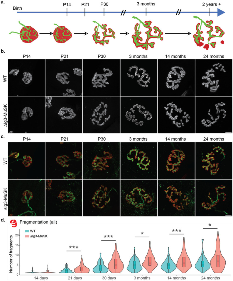
Figure 2. ΔIg3-MuSK NMJs exhibit increased postsynaptic fragmentation without anatomical denervation throughout the lifespan.
a. Schematic of structural changes observed during WT NMJ development and aging. Note perforation of postsynaptic apparatus between birth and P30 and further postsynaptic fragmentation during aging. b. Postsynaptic structure (α-bungarotoxin only) of NMJs from sternomastoid of P14, P21, P30, and 3-, 14-, and 24- month old WT and ΔIg3-MuSK mice, c. merged images of pre- and post- synaptic apparatus of NMJs in (b). Postsynaptic apparatus (α-bungarotoxin, red); presynaptic (neurofilament with VAChT or synaptophysin; all green). Note increased fragmentation of postsynaptic elements and their persistent co-localization with presynaptic elements. d. Quantification of ΔIg3-MuSK NMJ fragmentation throughout the lifespan. (* p<0.05, ** p<0.01, *** p<0.001, generalized linear models, full statistics presented in Tables 2-1 and 2-2, including median and interquartile range for each measurement, n for each age and sex, tests used, and results of statistical testing.)

