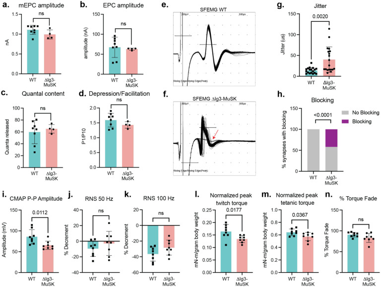
Figure 4. Impaired NMJ function in 6-month old ΔIg3-MuSK muscle.
a-d: Cholinergic signaling. Ex-vivo measurements of tibialis anterior (a.) NMJ spontaneous miniature endplate current (mEPC) amplitude, (b.) nerve-evoked endplate current (EPC) amplitude, (c.) quantal content, (d.) and depression/facilitation in response to trains of stimulation. All four parameters were indistinguishable between WT and ΔIg3-MuSK muscle. (n: a-d 4 mice/genotype (b: 2 WT)), 18–26 mEPCs, EPCs, or trains of stimulation). e, f: Representative waveforms of single fiber electromyography (SFEMG) recordings of muscle fiber action potentials (MFAPs) in WT (e.) and ΔIg3-MuSK (f.) gastrocnemius. Note the inconsistent latency of MFAPs and blocking in the ΔIg3-MuSK muscle (red arrow). Both jitter and blocking (quantified in g and h., respectively) were significantly increased in ΔIg3-MuSK (n = 3 mice/genotype, 5–7 trains of 50–100 stimuli per mouse; g. Student’s T-test, h. Chi-squared test.). i. Compound muscle action potential (CMAP) peak-to-peak amplitude is decreased in ΔIg3-MuSK (n: 6 mice/genotype, 1 stimulus per mouse), but CMAP % decrement in response to repetitive nerve stimulation (RNS) at (j.) 50 and (k.) 100 hz remains normal (n: 6 mice per genotype, 1 train of stimulus at each frequency per mouse). l. ΔIg3-MuSK mice produce less plantarflexion twitch torque in response to a single stimulus than WT. m. ΔIg3-MuSK mice produce less plantarflexion tetanic torque in response to a 1-second train of nerve stimuli than WT. n. Reduction of tetanic torque during a 1-s train of stimuli in ΔIg3-MuSK is similar to WT. (n: 6 mice/genotype, 1 twitch and 1 train of tetanic stimulus per mouse, Student’s t-test).

