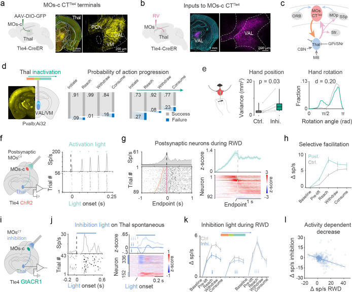
Fig 5. MOs-c CTTle4 selectively enhances RWD-relevant thalamic dynamics.
a. Neuron terminals of MOs-c CTTle4 in higher-order thalamus. Right: zoom-in view of the boxed region (middle). VAL, ventral anterior-lateral complex; VM, ventralmedial thalamus; PCN, paracentral nucleus.
b. Rabies (RV) tracing maps presynaptic inputs to MOs-c CTTle4 from VAL.
c. Schematics summary of the MOs-c corticothalamic reciprocal loop, which receives diverse cortical as well as subcortical inputs and projects to multiple cortical and striatal areas. Thal, thalamus; BG, basal ganglia, CBN, cerebellar nuclei; Str, striatum; OFC, orbitofrontal cortex.
d. Interference of thalamic dynamics during reach impairs RWD action progression. Left, ChR2 expression (yellow) and optic fiber implantation into VAL/VM; Right, 15% (78/511) control trials and 42% (201/482) inhibition trials failed to complete the RWD sequence. (n = 6 sessions from 4 mice)
e. Increased variance in hand position (left) and abnormal hand posture (right) upon lick with thalamic interference. (n = 6 sessions; Left: Wilcoxon rank sum test, . Right: two-sample KS test, .)
f. Electrophysiological recording of MOs-c CTTle4 postsynaptic neurons (TPNTle4-post) in thalamus. Blue light pulses were applied to ChR2-expressing CTTle4 neurons in the MOs-c (left). Light-evoked peri-event time histograms (top) and raster activity (bottom) of a TPNTle4-post (right). Note the facilitation of spiking with 20 Hz light pulses.
g. RWD related activity of individual TPNTle4-post. Left, spike raster of a TPNTle4-post during RWD. Right, summary of RWD related activity of 92 TPNTle4-post.
h. Increase in RWD-relevant activity of TPNTle4-post compared with control. ( TPNTle4-post and 210 control)
i. Electrophysiological recording from thalamus upon optogenetic inhibition of MOs-c CTTle4.
j. Effect of MOs-c CTTle4 inhibition (450 ms constant) on the spontaneous firing of thalamic neurons. Left: phasic decrease followed by brief increase of spontaneous discharge of a thalamus neuron during CTTle4 inhibition. Right, neurons were divided into decreased (Group I, 336), non-modulated (Group II, 326), and increased (Group III, 152).
k. Effect of MOs-c CTTle4 inhibition on RWD-relevant thalamus activity among the three groups. Inhibition light was delivered in random half of trials around lift during RWD.
l. Inhibition effect is dependent on RWD related increase in normal conditions. Each circle represents an individual Group I neuron. ( neurons,

