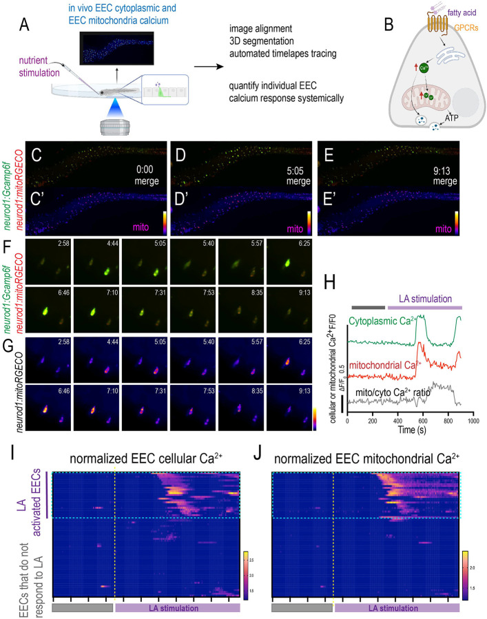
Figure 7. Analyze individual EEC cellular and mitochondrial activity in response to nutrient stimulation in live zebrafish.
(A) Image and analyze the EEC cellular and mitochondrial calcium activity before and after nutrient stimulation using the confocal microscope. (B) The hypothesis model figure supported by our experimental data shows fatty acid increases both cytoplasmic and mitochondrial Ca2+. Long-chain fatty acids, such as linoleic acid (LA), binds the fatty acid receptor at the cell membrane. Activating the fatty acid receptor induces Ca2+ release from the ER and increases the cytoplasmic Ca2+. The increased cytoplasmic Ca2+ is then translocated into the mitochondrial matrix, which increases the mitochondrial matrix’s Ca2+ levels. The increased mitochondrial Ca2+ promotes ATP production and powers EEC vesicle secretion. (C-E’) Time-lapse images of the whole zebrafish intestinal EECs’ cytoplasmic and mitochondrial calcium change post linoleic acid stimulation. The EEC cytoplasmic Ca2+ was labeled by Gcamp6f (green), and the EEC mitochondrial Ca2+ was labeled by mitoRGECO (red). (F-G) Zoom-out view shows two representative EECs that are activated by linoleic acid. (H) Analysis of fluorescence change of Gcamp, mitoRGECO, and mitoRGECO/Gcamp ratio in a presentative linoleic acid activated EEC. (I-J) Analysis of fluorescence change of Gcamp and mitoRGECO of 68 EECs in one zebrafish before and after linoleic acid stimulation. The EECs that increase cytoplasmic Ca2+ were defined as “LA activated EECs”. Noted that the majority of the linoleic activated EECs also exhibit increased mitochondrial calcium.

