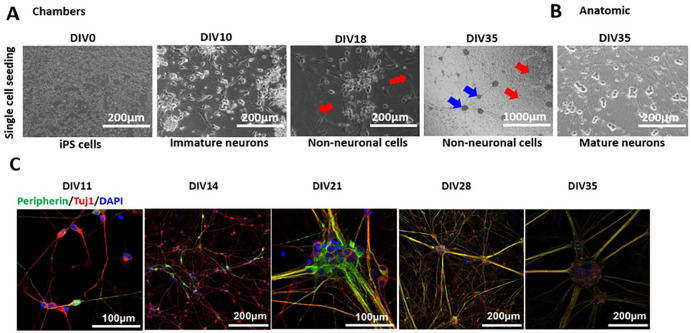
Figure 4. Chambers protocol: Differentiation and Maturation (Ctrl1).
A. Phase contrast images during the differentiation and maturation of iPSC-derived from Ctrl1 subject. DIV0 iPSCs, DIV10 generation of immature neurons, DIV35 shows ganglion-like morphology (marked with blue arrows). DIV18 and 35 also shows the culture outgrown with other non-neuronal cell types (marked with red arrows). Scale bar 1000μm and 200μm. B. Neurons at DIV35 from Anatomic protocol results in homogenous neuronal network. Scale bar 200μm. C. Immunostaining confirms peripheral neuronal identity of neurons from DIV11, 14, 21, 28 and 35 of maturation. DIV-Days in vitro. Scale bar 100 μm and 200μm. Peripherin-green, Tuj1-red and DAPI-blue fluorescence.

