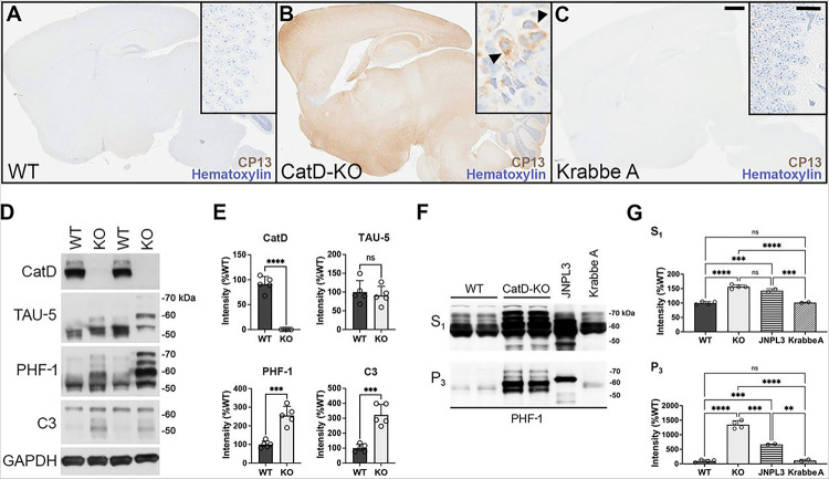
Figure 3. Three-week-old CatD-KO mice exhibit prominent, widespread tauopathy.
(A-C) Sagittal brain sections of wildtype (WT; A), CatD-KO (KO; B), and Krabbe A (C) mice stained for hematoxylin and the phospho-tau-specific antibody CP13. Note the abundant CP13 immunoreactivity evident in CatD-KO mice but lacking in WT or Krabbe A controls. High-magnification images of hippocampal CA3 regions (insets) reveal prominent perinuclear phospho-Tau staining in CatD-KO mice (B, arrowheads), resembling the localization of Gallyas silver staining seen in Figure 2. Scale bars (C) are 1 mm for all main images and 50 μm for all insets. (D,E) Western blotting (D) and quantitation thereof (E) in ~3-week-old WT and KO mice (n = 5 per genotype), showing levels of cathepsin D (CatD), total tau (TAU-5), phospho-tau (PHF-1) and caspase-cleaved tau (C3) along with GAPDH as a loading control. Note that total tau levels are not increased quantitatively (E, upper right graph), despite the pronounced perturbations in the migration pattern of total tau in CatD-KO mice consistent with hyperphosphorylation. (F,G) Representative PHF-1 western blots (F) and quantitation thereof (G) of soluble (S1) and sarkosyl insoluble (P3) brain extracts from ~3-week-old wildtype (WT) and CatD-KO mice (n = 4 per genotype) together with 9-month-old hTau transgenic (JNPL3) and 3-month-old Krabbe A controls (n = 2 per genotype). Note that CatD-KO mice harbor levels of sarkosyl-insoluble tau by just 3 weeks of age exceeding those present in 9-month-old JNPL3 mice, which show abundant tauopathy by this age [26]. Note further that Krabbe A mice, which feature marked lysosomal disturbances and were collected at a similar antemortem interval as the CatD-KO mice, show no increases in soluble or sarkosyl-insoluble tau relative to WT mice (F,G). Data are mean ± SEM; *P<0.05; **P<0.01; ****P<0.001; ****P<0.0001; ns=not significant.

