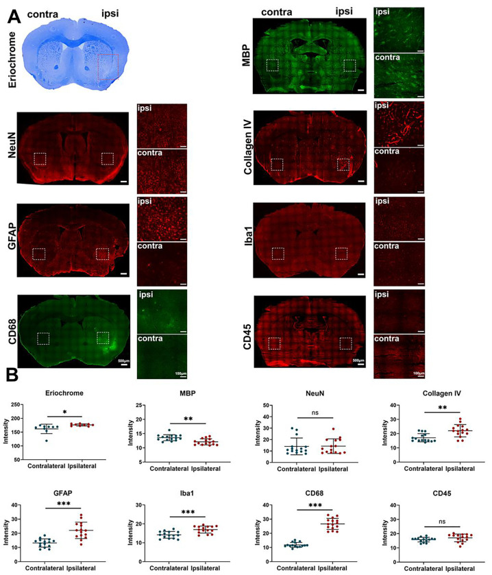
Figure 2. Histological evidence of brain damage after fractionated 3×20 Gy radiation.
(A) Representative histology/immunohistochemistry of brain tissue stained for Eriochrome, MBP, NeuN, Collagen IV, GFAP, Iba1, CD68, and CD45 at 80 weeks after fractionated 3×20 Gy radiation. The scale bars are 500 μm for low-power images and 100 μm for high-power images. (B) Quantification of signal intensity in the contralateral and ipsilateral hemispheres for each staining. Paired t-test. N=3 ROIs from 3 (Eriochrome staining) or 5 (all the immunofluorescence staining) brain slices in each group. *P<0.05, **P<0.01, ***P<0.001. ns: no significant difference.

