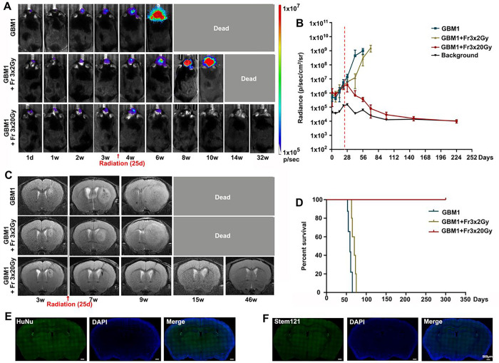
Figure 3. Eradication of GBM1 tumor with high-dose radiotherapy.
(A) Representative BLI images of non-irradiation, fractionated 3×2 Gy or 3×20 Gy irradiation-treated tumor-bearing mice at the indicated days. Irradiation was started on day 25 after tumor inoculation. (B) Quantification of the BLI signal revealed that 3×20 Gy radiation completely eradicates the GBM1 tumor. The dot-dashed line represents the date of starting radiation. (C) Representative T2-weighted MR images for GBM1 mice without radiation or with fractionated 3×2 Gy or 3×20 Gy radiation. (D) Survival curves of GBM1 mice with or without radiation. N=5 mice/group. (E) HuNu staining. (F) Stem 121 staining. The scale bars are 500 μm.

