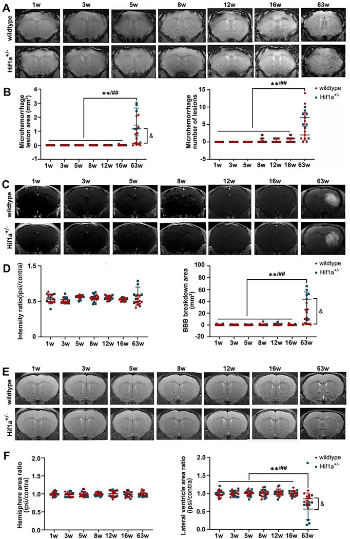
Figure 4. Under longitudinal MRI, monitoring brain damage after fractionated 3×20 Gy radiation in both HIF-1α+/− heterozygote and wild-type mice.
(A-B) Representative T2* images and microhemorrhage assessment for mice receiving fractionated radiation. (C-D) Representative T1-weighted gadolinium-enhanced MRI and BBB status for mice receiving fractionated radiation. (E-F) Representative T2-weighted MR images and anatomical analysis for mice receiving fractionated radiation. N=3 mice per group (3 slices for each mouse). Wildtype group: */**P 0.05/0.01. HIF-1α+/− group: #/##P 0.05/0.01. wildtype vs. HIF-1α+/− group: &P 0.05.

