
Figure 4.
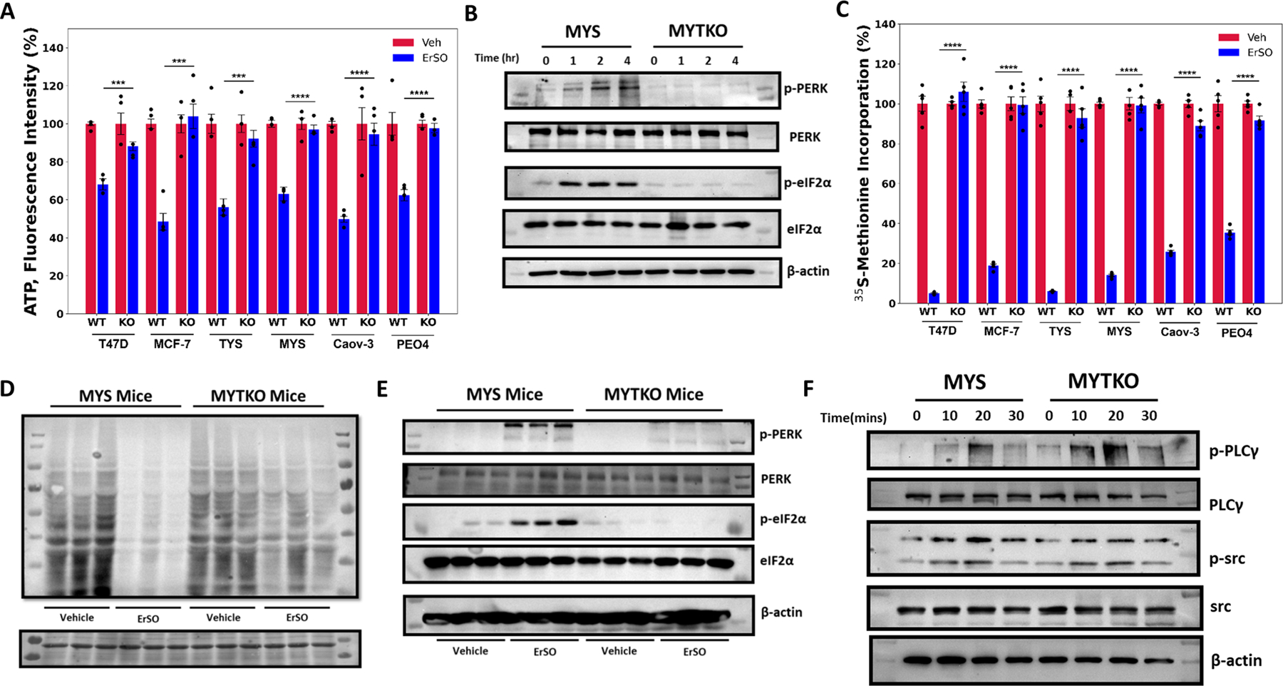
TRPM4 sustains lethal UPR hyperactivation. A Whole cell ATP levels of the indicated wild type and TRPM4 knockout breast and ovarian cancer cells treated with vehicle (set to 100%) or 1 μM ErSO for 4 hours (n=4). B Western blot analysis of p-PERK, total PERK, p-eIF2α, total eIF2α and β-actin in MYS cells and MYTKO cells at 0, 1, 2 and 4 hours after treatment with 1μM ErSO. C Incorporation of 35S-methionine into newly synthesized protein in the indicated wild type and TRPM4 knockout breast and ovarian cancer cell lines treated with vehicle (set to 100%) or 1 μM ErSO for 1 hour (n=5). D, E MYS-Luc and MYTKO-Luc cells were orthotopically grafted in NSG mice and tumors allowed to grow for 21 days. Mice were then treated with either vehicle or ErSO (40 mg/kg i.p) for 5 hours (n = 3). D Mice were then injected with puromycin in sterile PBS (0.04 μmol/ per g of body weight). After 1 hour, mice were euthanized and tumors were resected. Protein lysates from the tumors were prepared and puromycin-labeled peptides were identified using Western blot analysis. Ponceau S staining was used as a loading control (on the bottom). E Mice were euthanized and tumors were resected. Protein lysates from the tumors were prepared and Western blot analysis was performed for p-PERK, total PERK, p-eIF2α, total eIF2α and β-actin. F Western blot analysis of p-PLCγ, total PLCγ, p-src, total src and β-actin in wild type MYS cells and MYTKO cells at 0, 10, 20, and 30 minutes after addition of 1 μM ErSO. Data are mean ± s.e.m. ***p<0.001, ****p<0.0001.