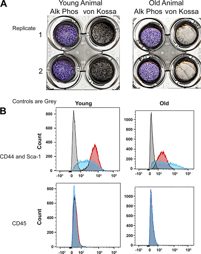
Figure 2.
Characterization of stromal stem cells and osteoblasts. A: examples of alkaline phosphatase and von Kossa stains for 2 replicates out of 4 young (2 males and 2 females) and 4 old (2 males and 2 females) mice. Each well is a whole well from a 6-well plate with the inner part of the image the perforated polyethylene terephthalate membrane containing the complete ∼1.6-cm wide cell culture. The images shown are from 3-wk differentiated cultures, where transepithelial resistance is ∼1,400 Ω-cm (9). B: flow cytometry showing that preparations essentially uniformly expressed CD44 and Sca1, but not CD45 (negative control). Gray bars are isotype controls. Example are shown; there was no meaningful differences in flow analysis of any SSC culture out of 4 young (2 males and 2 females) and 4 old (2 males and 2 females) mice. SSC, stromal stem cell.

