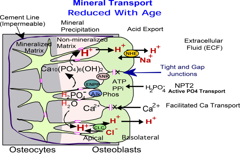
Figure 6.
Hypothetical model of bone formation pathway in osteoblasts. This model is for bone formation only and does not include bone degradation, not studied. Our results indicate that age-dependent decline in trabecular bone correlates with downregulation of expression of major bone matrix proteins, collagen 1 and osteocalcin, together with PPi-related alkaline phosphatase (ALP), ecto-nucleotide pyrophosphatase/phosphodiesterase (ENPP2), ankylosis (ANK) proteins, and Cl−/H+ antiporter ClC3. These proteins are located on the apical (bone forming) side of the epithelial-like layer of osteoblasts connected by tight and gap junctions and are responsible for the mineral and ion transport between cytoplasm and extracellular matrix. This is hypothetical in the case of ClC3, better known as an endosomal protein. Basolateral proteins, on the extracellular fluid side of the osteoblast epithelium-like layer (NPT2 and NHE1), appear not to be significantly regulated with age.

