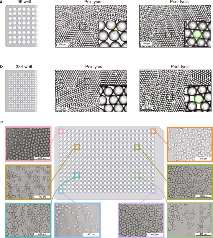
Extended Data Fig. 2. High cell and sample number experiments with PIP-seq.
(a-c) Microscope images of droplets and cells in plate emulsification experiments. Representative images are from experiments completed at least three times. (a,b) Barcode bead templates, stained cells (puncta), and lysis reagents are combined with oil and vortexed in (a) 96-well and (b) 384-well plates to generate monodispersed droplets. Heat activation of Proteinase K results in lysis and release of calcein dye, and full-drop fluorescence. (c) Microscope images of droplets from random wells of a 384-well plate emulsification experiment.

