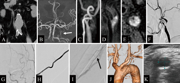
Fig. 1.
A) Computed tomography (CT) shows an abdominal aortic aneurysm, 44 mm in diameter. B) Magnetic resonance angiography of the head reveals a decreased signal from the left intracranial internal carotid artery (ICA) due to severe stenosis (white arrow). C) Time-of-flight magnetic resonance imaging demonstrates a high-intensity signal of the common carotid artery (CCA). D) Magnetization-prepared rapid gradient echo imaging shows a high-intensity signal in the CCA. E) T1-weighted black blood imaging reveals a high-intensity signal from the CCA, suggesting a vulnerable plaque. F) Left CCA angiography (lateral view) shows anterograde string-like filling of the ICA beyond the carotid bifurcation. G) Left CCA injection (frontal view) demonstrates prolonged cerebral circulation time in the left cerebral hemisphere. H) Contrast-enhanced CT angiography shows a bovine-type arch of the aorta. I, J) Right brachial angiography demonstrates a well-developed deep brachial artery (black arrow). K) Vascular echocardiography shows that the right brachial artery at the cubital fossa has a vascular diameter of approximately 4.7 mm.

