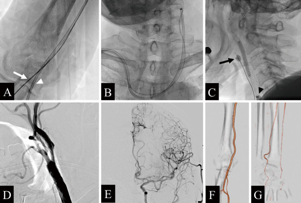
Fig. 2.

Angiography during carotid artery stenting. A) An 8-Fr balloon guide catheter (BGC) is navigated from the right brachial artery (white arrowhead), and a 4-Fr catheter is guided from the right radial artery (white arrow). B) BGC and 4-Fr catheter navigated to the left common carotid artery (CCA). C) Fluoroscopic lateral view in which postdilation has been performed after placement of the carotid stent using a flow-reversal system with a microballoon catheter (MBC) (black arrow) and a BGC (black arrowhead). D) Left CCA injection (lateral view) after all procedures were completed. E) Left CCA angiography (frontal view) reveals no contrast-enhanced defect. F, G) Contrast-enhanced computed tomography angiography after 5 days of intervention shows no puncture site complications in the right brachial (F) and radial (G) arteries.
