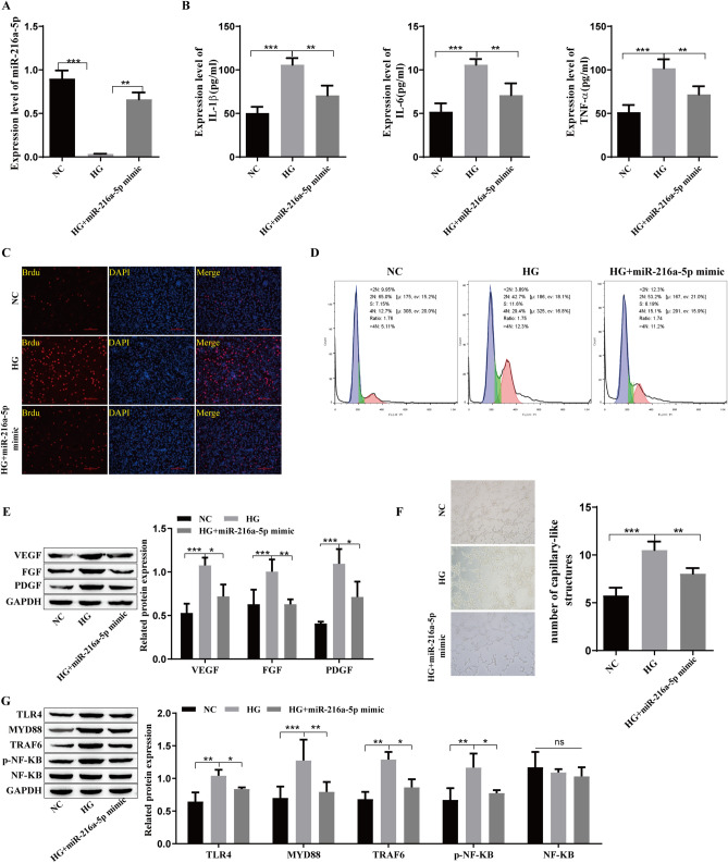
Fig. 2.
Influence of miR-216a-5p overexpression on inflammatory cytokines and growth factors in retinal microvascular endothelial cells. A: qRT‒PCR analysis of the level of miR-216a-5p. B: The expression of the inflammatory cytokines TNF-α, IL-6 and IL-1β was detected by ELISA. C: BrdU staining to detect cell proliferation. D: Flow cytometry to detect the cell cycle. E: Western blot analysis of the expression levels of the growth factors VEGF, FGF and PDGF. F: Angiogenesis was detected by tubule formation experiments. G: Western blot analysis of the level of TLR4/NF-kB-related proteins such as TLR4, MYD88, TRAF6, p-NF-kB and NF-kB. *P<0.05, **P<0.01, ***P<0.001

