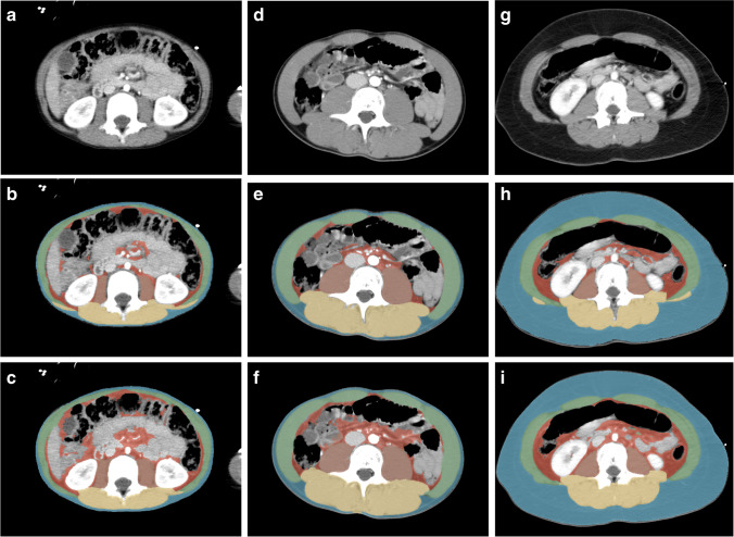
Fig. 1.
Axial computed tomography images obtained in a 5-year-old boy (a–c), a 15-year-old boy (d–f) and a 12-year-old girl (g–i) at the level of the third lumbar vertebral body performed following high-energy trauma. Contrast-enhanced images before segmentation (a, d, g) and after manual (b, e, h) and automatic (c, f, i) segmentation. Automatic segmentation sometimes included parts of the intestines as visceral fat and often failed to differentiate the abdominal wall muscles from the latissimus dorsi muscles. Blue subcutaneous fat, green abdominal wall muscles, red visceral fat, orange psoas muscles, yellow paraspinal muscles

