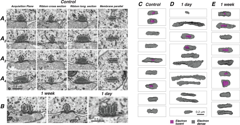
Figure 3.
Pre-synaptic ribbons hypertrophy and duplicate after synaptopathic noise. (A1–A4) The apparent heterogeneity of ribbon morphology disappears when each synapse is virtually re-sectioned in a ribbon-centric plane, as enabled by the isotropism of FIB-SEM image stacks. Four examples from a control ear appear different from each other when viewed in the acquisition plane. The same four synapses look similar to each other when viewed either as either (2nd column)—ribbon cross-section, i.e. perpendicular to the subjacent IHC membrane and to the long axis of the ribbon; (3rd column)—ribbon longitudinal section, i.e. perpendicular to the membrane and parallel to the long axis of the ribbon; or (4th column)—membrane parallel to the adjacent IHC and through the middle of ribbon thickness. (B) Double and triple ribbons are more common in noise-exposed ears, at either 1-week or 1-day survival as indicated. (C–E) The post-exposure hetererogeneity of ribbon length and number is illustrated by this sampler of ribbon morphologies from control, 1-day and 1-week cases, respectively. For each survival time, the sample include the longest and shortest examples from the group. Each box outlines the ribbon(s) at a different ANF synapse. Each image is a 3D reconstruction of the ribbon volume, viewed perpendicular to the plane of subjacent IHC membrane, i.e. in the same orientation as the membrane parallel images from panels (A1–A4). Each surface is rendered with enough transparency to see the hollow core (purple) when present. When multiple ribbons are present, their relative orientations are maintained.

