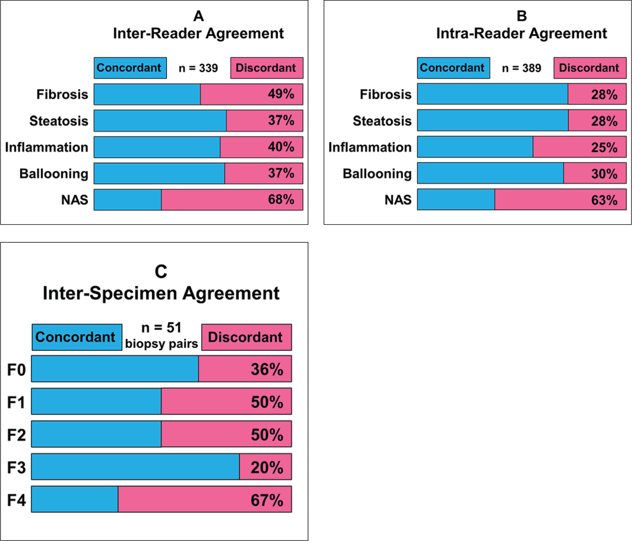
Figure 2.

Metrics showing limitations in diagnostic performance of biopsy for evaluation of liver fibrosis, as reported in literature.
A-In study in which 339 liver biopsy specimens were each evaluated independently by two expert histopathologists, reported stage of fibrosis in specimens was discordant in nearly half of specimens. Substantial discordance was also present for grading of steatosis, inflammation, ballooning, and assignment of nonalcoholic fatty liver disease activity score (NAS) [9}.
B-In same study, 389 liver biopsy specimens were each evaluated at two separate times by same expert histopathologist. Reported severity of fibrosis and other histopathologic findings for same specimens were discordant in 25–63% of cases [9].
C-In study that obtained two percutaneous liver biopsy specimens from each of 51 patients, stage of fibrosis in pairs of specimens evaluated by expert histopathologist was discordant in 20–67% of cases [12].
