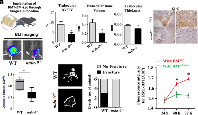
Fig. 2.
Lack of MDA-9/Syntenin expression inhibits tumor outgrowth in the bone niche. (A) RM1-BM-Luc cells were directly implanted (into the femur) in 2-mo-old WT and mda-9−/−mice (Top panel: schematic presentation). Representative images at day 14 are presented (Middle). Luciferase intensity was calculated using built-in Living Image Software v.2.50, and the average value for each group at day 14 is graphically presented (Bottom). Error bars ± SD. (B) Quantification of mineralized tissue by µCT in the distal femur. Femurs were assessed for amount of trabecular bone volume over total volume, bone volume alone, and thickness of the trabeculae in the distal femur. (C) Prevalence of pathologic fracture as observed by µCT. (D) Tumor-implanted bones (from animals studied in panel A) were immunostained for KI-67 expression, and the representative photographs at lower (Left) and higher magnifications (Right) are presented. (E) Bone marrow cells from WT (BMWT) and mda-9−/− (BMmda-9−/−) mice were isolated and cocultured with fluorescently labeled tumor cells (RM1-BM-Luc) and incubated for different time periods. FACS analyses were performed to detect tumor cells at different time points. *Statistically significant (P < 0.05).

