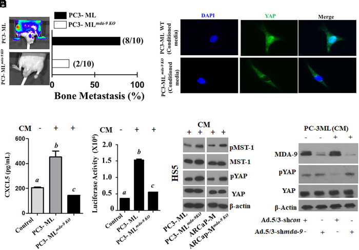
Fig. 4.
MDA-9 expression in tumor cells nonautonomously activates the Hippo pathway and induces CXCL5 in HS5 cells. (A) Bone metastasis development in athymic nude mice following intracardiac injection of PC-3ML control and mda-9 KO (PC-3MLmda-9 KO) clone (1 × 10⁵ cells/mouse). Representative images 36 d after tumor cell implantation (Left). Bone metastases detected by BLI imaging. The percentage of mice with BLI-positive lesions is shown (Right). (B) HS5 cells were incubated with normalized (equal amount of total protein) tumor cell–derived condition media for 12 h. HS5-derived conditioned media were analyzed for CXCL5 levels using ELISA. (C) HS5 cells were transfected with a 1.5-kb CXCL5-Prom using a standard transfection protocol. Twelve hours posttransfection, cells were stimulated with conditioned media for an additional 12 h. Luciferase activity was measured and presented after normalizing with Renilla luciferase. (D) HS5 cells were incubated with tumor cell–derived conditioned media for 12 h and stained with fluorescently labeled anti-YAP. DAPI was used for nuclear staining. Translocation of YAP into the nucleus was monitored under a fluorescence microscope. (E) HS5 cells were incubated with tumor cell– (parental and its mda-9 knockout variant) derived conditioned media for 12 h, and cell lysates were analyzed for different proteins, as indicated. (F) MDA-9/Syntenin expression was knocked down in HS5 cells using an Adenovirus expressing shmda-9. Twenty-four hours postinfection, culture media were replaced with fresh media containing PC-3ML CM for an additional 12 h. Cell lysates were analyzed for the indicated proteins. Different letters in two variables are statistically significant (P < 0.05).

