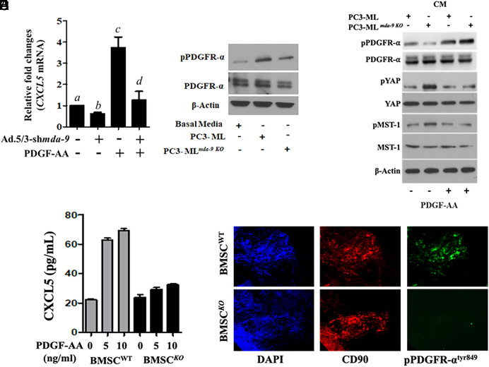
Fig. 7.
MDA-9 expression in HS5 regulates PDGF/PDGFRα signaling. (A) mda-9 expression was blunted, and 24 h postinfection, HS5 cells were stimulated with PDGF-AA for an additional 12 h. mRNA for CXCL5 expression was analyzed using qPCR. Different letters in two variables are statistically significant (P < 0.05). (B) HS5 cells were incubated with media supplemented with the indicated tumor cell–derived conditioned media for 12 h, and western blotting was done for phosho-PDGFRα and total PDGFR. (C) HS5 cells were incubated with media supplemented with the indicated tumor cell–derived conditioned media and PDGF-AA for 12 h, and western blotting was done for the indicated proteins. (D) BM-MSCs from WT and mda-9−/− mice were stimulated with mouse PDGF-AA, and CXCL5 expression was determined in the culture media. (E) phospho-PDGFR expression was analyzed in paraffin sections (tumor-bearing bone from the animal experiment, described in Fig. 2A). Different letters in two variables are statistically significant (P < 0.05).

