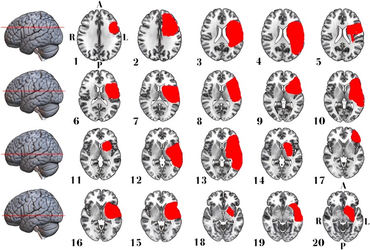
Figure 4.
Stroke lesion distribution in patients with aphasia. This figure displays 2D maps of traced ischaemic stroke or haemorrhage lesions in radiological convention, each representing a separate patient. Lesions led to aphasia in 20 patients (65% women, median age of 73 years). All lesions were in the left hemisphere, and most were mixed cortical/subcortical. Directional abbreviations indicate brain orientation: A, anterior; L, left; P, posterior; R, right.

