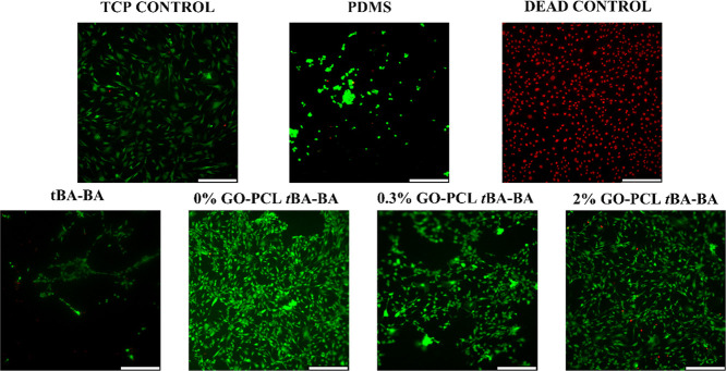
Figure 7.
Live/Dead micrographs of C3H10T1/2 cells taken after triple-shape recovery showing high sample cytocompatibility. Few dead cells (red dots) were observed in any of the groups, except for the dead control. TCP (tissue culture plate), PDMS (polydimethylsiloxane), tBA-BA (tert-butyl acrylate-butyl acrylate), GO (methacrylated graphene oxide), and PCL (poly(ε-caprolactone)). Scale bar: 330 μm.

