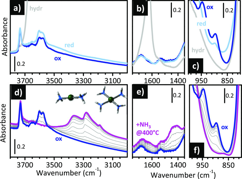
Figure 13.
IR spectroscopy is sensitive to the oxidation state and local environment of the Cu sites in Cu-SSZ-13 zeolite. (a–c) IR spectroscopy allows the speciation and quantification of the different Cu sites in Cu-exchanged zeolites. (a–c) IR spectra of Cu-SSZ-13 in its hydrated form (hydr), after thermal treatment in O2 at 400 °C (ox), and after prolonged thermal treatment in inert flow at 400 °C (red) in three different spectral regions. (d–f): IR spectrum of Cu-SSZ-13 after thermal treatment in O2 at 400 °C (ox), and its time evolution during interaction with NH3 at 400 °C. The pink spectrum was collected after 30 min. The insets in (d) show the structure of linear diammino and square-planar tetraammino Cu complexes formed in the presence of NH3 at 400 °C. Unpublished data.

