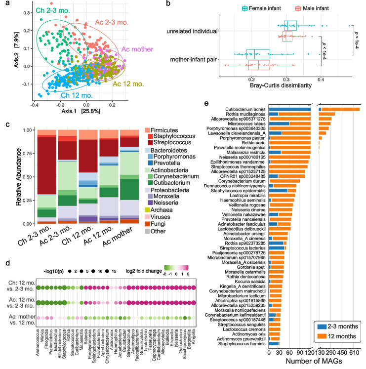
Fig. 3.
Early-life skin microbial community structure. a Principal coordinate analysis (PCoA) on Bray–Curtis dissimilarity between the microbial profiles. Each point represents a single sample and is colored by body site and age group. Ellipses represent a 95% confidence interval around the centroid of each sample group. b The Bray–Curtis dissimilarity of mother–infant pairs comparing related versus unrelated dyads. Median value of each infant and all unrelated mothers was used. Statistical difference was tested by two-sided Wilcoxon rank sum test. c Relative abundance of skin microbiome averaged for each sample group. Two of the most abundant genera within each bacterial phylum were shown. d Differential taxa at the genus level between infants of different ages and between infants at 12 months and mothers. The size of the dots represents the log-transformed adjusted p-value from DESeq2, and the color indicates fold changes. The top differentially abundant genera for each comparison were shown. e Number of species-level MAGs recovered from infants at 2–3 months and 12 months, sorted by the total number of MAGs

