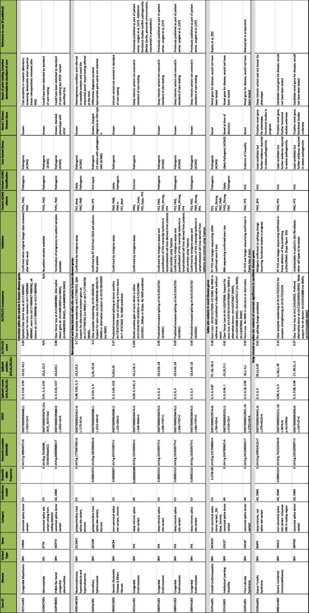
Table 2.
Splice site and deep intronic variants identified in the OxClinWGS cohort
The table is divided into four sections, according to whether variants are (i) canonical splice site variants in known genes (n = 3); (ii) non-canonical splice site variants in known genes (n = 7, of which 5 are unique); (iii) splice site variants in novel (n = 2), or putative novel (n = 1) disease genes; and (iv) variants of uncertain significance in candidate genes (n = 3). Sixteen splice site variants (fourteen unique variants) were identified in fifteen cases. Thirteen variants contributed to cases which we considered to be solved, as the variants were classified as pathogenic/likely pathogenic (according to ACMG criteria) or were clinically actioned. Ten variants were in known genes for the phenotype, although seven of these were either non-canonical splice sites or deeper into introns and would not have been detected by conventional testing. Two splice site variants were in novel genes (or genes that were novel at the time of discovery) whilst one is in a novel gene with evidence of causality. Three were variants of uncertain significance, two of which were in the same patient which could account for different elements of the patient’s phenotype. We defined a canonical splice site variant as ± 2 bp into intron
Details of prior testing and whether variants could have been detected by the standard of care testing (arrays, panels or exomes) are included
Abbreviations: AR autosomal recessive, AD autosomal dominant, CH compound heterozygous, DNM de novo mutation, CDA congenital dyserythropoietic anaemia type II

