
Figure 2.
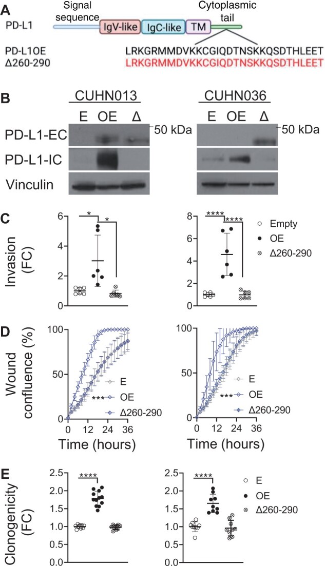
Programmed death ligand 1 (PD-L1) intracellular domain and intrinsic function. A) Schemata of PD-L1 structural domains. PD-L1 intracellular domain (PD-L1–ICD) is expanded to amino acid structure below. PD-L1 overexpression and PD-L1–ICD deletion mutant (Δ260-290PD-L1) shown below. Created with BioRender.com. B) Confirmation of Δ260-290PD-L1 overexpression in CUHN013 and CUHN036 by immunoblot. C) PD-L1 overexpression increases invasion, (D) wound healing, and (E) clonogenic growth, but this is reversed with Δ260-290PD-L1 overexpression (wound confluence 16 hours postscratch: CUHN013 empty = 46.59%; PD-L1 overexpression = 85.59%; Δ260-290PD-L1 = 47.11%; CUHN036 empty = 46.59%; PD-L1 overexpression = 87.51%; Δ260-290PD-L1 = 51.20%). *P ≤ .05, **P ≤ .01, ***P ≤ .001, ****P ≤ .0001. Error bars represent standard deviation. E = empty; FC = fold change; IgC = immunoglobulin constant; IgV = immunoglobulin variable; OE = PD-L1 overexpression; Δ and Δ260-290 = Δ260-290PD-L1 overexpression; PD-L1-EC = PD-L1 extracellular-specific antibody; PD-L1-IC = PD-L1 intracellular specific antibody.