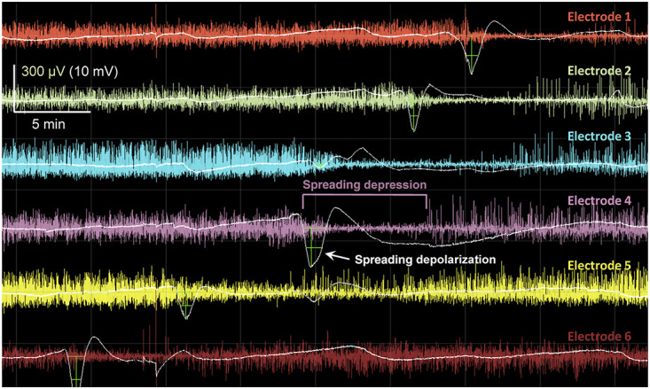
FIGURE 1.

Spreading depolarizations in electrocorticographic recordings of severe brain trauma. Illustration of a spreading depolarization propagating along a 6-contact subdural electrode strip, sequentially from electrodes 6 to 1. For each electrode, the raw recording is filtered for the typical electroencephalography band of 0.5 to 50 Hz (color traces) and the slow potential recording (<0.1 Hz) is overlaid (white traces; negative is down). The slow potentials are filtered by subtraction of the 30-minute moving median to correct for baseline drift. Green crosshairs mark the negative direct current shifts that are characteristic of spreading depolarizations. The sustained (typically 1-3 minutes) mass depolarization of cortex precludes electrical signaling, resulting in a spreading depression of the amplitude of electroencephalography band activity.
