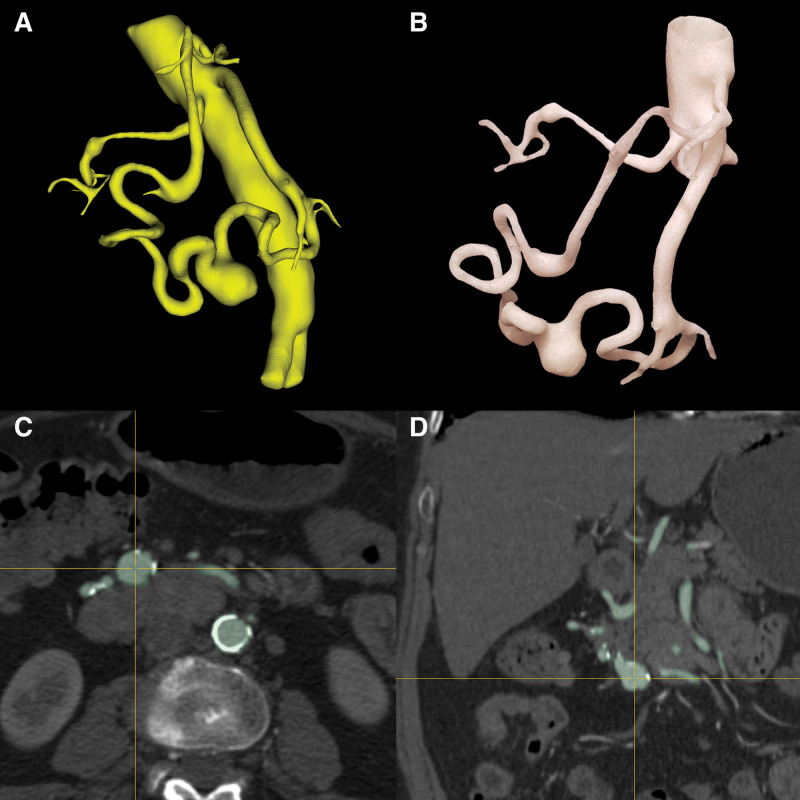
Figure 4.
The printed 3D model on the 1:1 scale (B) enables the accurate assessment of many changes without other anatomical structures visible in the angio-CT examination (C, D). In the figures, dolichoectasia and an aneurysm in the course of the upper pancreatic-duodenal artery and the lower pancreatic-duodenal artery with additional RRAA are shown. Virtual model (A). RRAA = right renal artery aneurysm.

