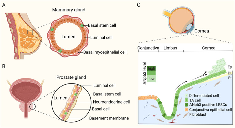
Fig. 3. Stem cell in glandular and stratified epithelia.
A A schematic model depicting the structure of the mammary gland epithelium, composed by basal stem cells, basal myoepithelial cells, and luminal cells. B The model shows the distribution of basal stem cells, luminal cells, and neuroendocrine cells in the prostate gland epithelium. C Schematic representation of the cornea. LESCs produce daughter TA cells that migrate towards the apical layer of the central cornea, and, eventually, become terminally differentiated (arrowed). ΔNp63 is the dominant p63 isoform expressed in LESCs and its expression decreases from limbus to the central cornea. Ep epithelium, BL Bowman’s layer, St stroma.

