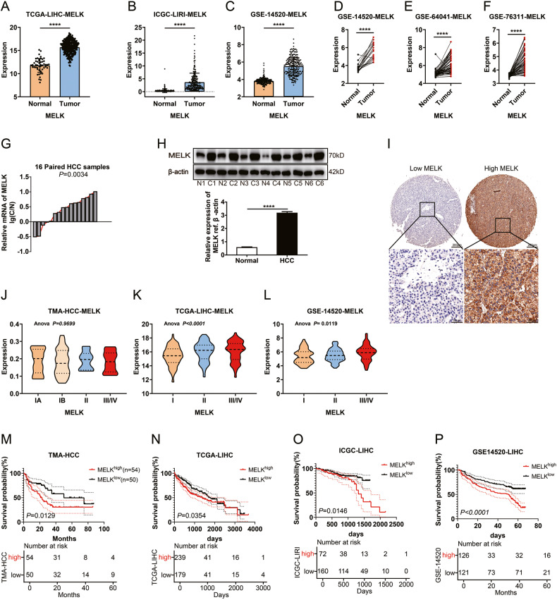
Fig. 1. Expression and clinical significance of MELK in HCC.
A–F Amplification of MELK is common in TCGA and GEO provisional HCC cohorts. The expression of MELK was detected with qPCR in 16 pairs of HCCs and adjacent tissues (G) and with WB in six randomly-selected pairs of HCC tissues (H). I Representative images of immunohistochemical staining for low/high expression of MELK in the tissue microarray (top: ×100 magnification, scale bar, 200 μm; bottom: ×400 magnification, scale bar, 50 μm). The expression of MELK in HCC patients with different clinical stages in TMA (J), TCGA (K), and GEO (L) provisional HCC cohort. The prognostic significance of MELK in TMA (M), TCGA-LIHC (N), ICGC-LIRI (O), and GSE14520 (P) cohorts. The p values were analyzed by Student’s t test and one-way or two-way ANOVA. The Kaplan–Meier method was used for prognosis analysis. Data were from at least three independent experiments and shown as the mean ± SEM ****P < 0.0001.

