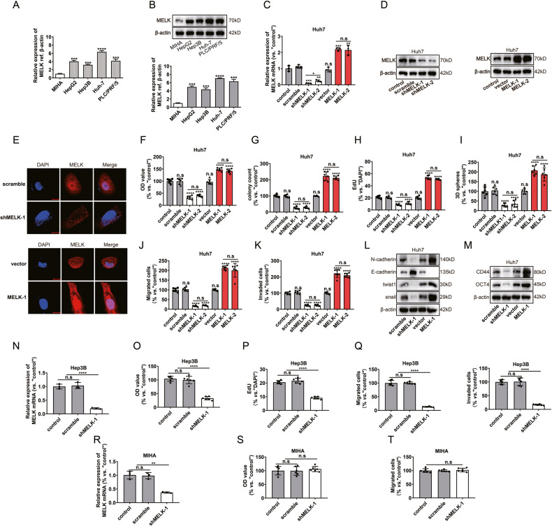
Fig. 2. MELK promoted HCC cell progression in vitro.
A, B The mRNA and protein expressions of MELK in normal human hepatocytes (MIHA) and different HCC cell lines: HepG 2, Hep 3B, Huh7, and PLC/PRF/5. After MELK expression was silenced or overexpressed in Huh7 cells: MELK expression was detected by qPCR (C), WB (D) and immunofluorescence (E); Cell proliferation was evaluated with CCK-8 assay (F), colony formation assay (G) and EdU assay (H); Stemness was evaluated with the 3D sphere formation assay (I); Cell migration and invasion were investigated with transwell assays (J, K); Biomarkers of EMT and stemness were detected by WB (L, M). After MELK knockdown in the HCC cell line Hep3B and the normal human hepatocytes MIHA: MELK expression was detected by qPCR (N, R); Cell proliferation was evaluated with CCK-8 assay (O, S) and EdU assay (P); Cell migration and invasion were investigated with transwell assays (Q, T). Data were shown as mean ± SEM. n.s. represents not significant. *P < 0.05, **P < 0.01, ***P < 0.001 and ****P < 0.0001 as calculated by the one-way or two-way ANOVA. Scale bar: 10 µm.

