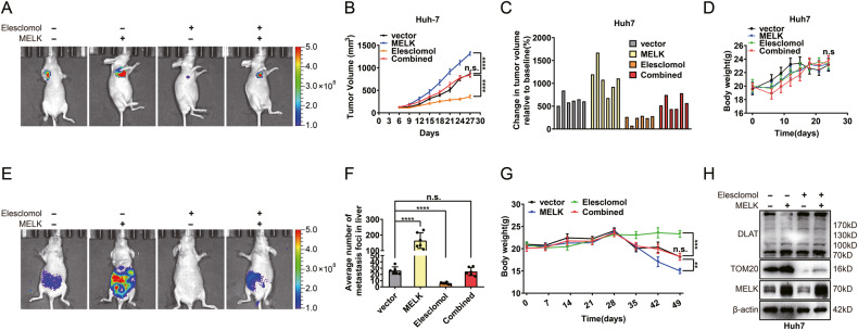
Fig. 7. The cuproptosis-associated pathway was essential in MELK-induced HCC progression in vivo.
A Xenograft models were established by subcutaneous injection of MELK-overexpressing Huh7 cells, elesclomol (10 mg/kg s.c.) was used to regulate DLAT status in vivo. Changes in subcutaneous xenograft tumors and body weight were observed (B–D). E Metastatic models were established by tail vein injection of MELK-overexpressing Huh7 cells, elesclomol (10 mg/kg s.c.) was used to regulate DLAT status in vivo. The tumor metastases were monitored by a live imaging system. The metastatic nodules in the liver were measured (F). And the changes in body weight of indicated groups of nude mice models were observed (G). H The expression of TOM20 and DLAT of tumors assessed via WB. Data were shown as mean ± SEM. n.s. represents not significant. *P < 0.05, **P < 0.01, ***P < 0.001 and ****P < 0.0001 as calculated by the one-way or two-way ANOVA.

